University students with subthreshold depression, meaning real depressive symptoms that fall short of a major depression diagnosis, had stronger connections in 3 cingulate brain circuits and slightly higher levels of 3 inflammatory proteins than healthy students, according to a 2026 brain-imaging study from China.1
Research Highlights
- Stronger cingulate connections: resting brain scans of 126 students with subthreshold depression and 104 healthy students showed stronger links between parts of the cingulate cortex and the caudate, the cerebellum and the superior frontal gyrus. No circuit was weaker.1
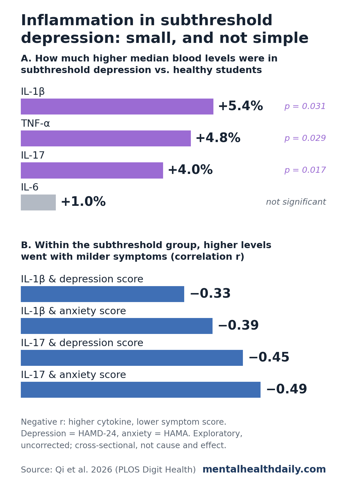
- Slightly higher inflammation: in the 125 students with blood results, median IL-1β, IL-17 and TNF-α were about 4% to 5% higher in the subthreshold group (p = 0.017 to 0.031). IL-6 did not differ.1
- An unexpected twist: within the subthreshold group, students with higher IL-1β and IL-17 had milder depression, anxiety and rumination scores.1
- No direct cytokine-brain correlation: cytokine levels did not correlate with any of the altered connections. They were linked to anxiety and rumination only through interaction terms in regression models.1
- Early-stage evidence: the study was cross-sectional and exploratory, with uncorrected symptom analyses and blood data missing for 46% of participants.1
What Subthreshold Depression Is and Why It Gets Studied
Subthreshold depression describes people who have at least one core symptom of depression, such as low mood or loss of interest, along with some trouble functioning, but who do not meet the full criteria for major depressive disorder. It also goes by minor, subclinical or subsyndromal depression.1
It is more common than major depression and easy to overlook because the symptoms are milder. It also carries real risk: a 2019 meta-analysis of long-term cohort studies found that people with subthreshold depression developed major depression at about 2 times the rate of people without depressive symptoms (incidence rate ratio 1.95).2
That risk is why researchers look for biological markers at this stage. If a measurable brain or blood signature could flag who is most likely to get worse, prevention could be aimed more precisely.
How the Study Measured Brain Connectivity and Inflammation
Zhangzhang Qi et al. at Jinan University in Guangzhou recruited university students between 2019 and 2023 and published the results in PLOS Digital Health.1
- Subthreshold group: 126 students aged 18 to 35 who scored 16 or higher on the CES-D depression questionnaire and 8 to 20 on the 24-item Hamilton Depression Rating Scale (mild range), with no major depression diagnosis and no antidepressants in the past year
- Comparison group: 104 healthy students who scored below both cutoffs
- Who was excluded: anyone with another psychiatric diagnosis, recent infection, allergy, autoimmune disease, obesity, current smoking or alcohol abuse (several of these raise inflammation on their own)
- Symptoms: depression (CES-D and Hamilton depression scale), anxiety (Hamilton Anxiety Scale) and rumination (Ruminative Responses Scale)
On average, the subthreshold group scored 14.1 on the Hamilton depression scale vs. 2.1 for the healthy students, and 14.7 vs. 1.4 on the anxiety scale. Age, sex and years of education were similar.1
Functional connectivity is a measure of how closely the activity of 2 brain regions rises and falls together while a person lies quietly in an MRI scanner. Regions with high connectivity are thought to be working as a team. It does not show physical wiring or which region drives the other.
The researchers focused on the cingulate cortex, a curved band of tissue on the inner surface of each hemisphere, just above the bundle of fibers that joins the 2 sides of the brain. It helps combine emotion with attention and self-control, and it has been implicated in depression for decades.
The team split the cingulate into 5 parts on each side and mapped each part’s connections across the whole brain.1
Cytokines are small signaling proteins the immune system uses to coordinate inflammation. The team measured 4 pro-inflammatory cytokines in fasting morning blood samples: interleukin-1 beta (IL-1β), interleukin-6 (IL-6), interleukin-17 (IL-17) and tumor necrosis factor alpha (TNF-α).1
3 Cingulate Brain Circuits Were More Connected in Subthreshold Depression
Compared with healthy students, the subthreshold group showed stronger connectivity in 3 circuits:1
- Middle cingulate to caudate: the caudate is part of the basal ganglia, deep structures involved in reward, motivation and habits. Stronger connectivity here went with higher CES-D depression scores (r = 0.19) and more rumination (r = 0.19).
- Front-upper cingulate to cerebellum: the left supracallosal anterior cingulate (the front part sitting above the fiber bundle) was more connected to the front of the cerebellum. The cerebellum is best known for balance and coordination but also contributes to emotion and thinking.
- Front-upper cingulate to superior frontal gyrus: on the right side, the same cingulate region was more connected to the superior frontal gyrus, part of the prefrontal cortex involved in planning and regulating emotion. Stronger connectivity here went with higher anxiety scores (r = 0.25).
No cingulate circuit was weaker in the subthreshold group. The symptom correlations were small, and the researchers did not correct them for the number of tests run.1
IL-1β, IL-17 and TNF-α Were Slightly Higher; IL-6 Was Not
Usable blood results were available for only 75 students with subthreshold depression and 50 healthy students. The other 105 samples were lost to non-cooperation or too little serum, broken red blood cells in the sample, or assay failure.1
In those 125 students, the subthreshold group had higher levels of 3 cytokines:1
- IL-1β: median 5.83 vs. 5.53 pg/ml (p = 0.031)
- TNF-α: median 130.25 vs. 124.30 pg/ml (p = 0.029)
- IL-17: median 18.95 vs. 18.22 pg/ml (p = 0.017)
- IL-6: median 12.27 vs. 12.15 pg/ml, no meaningful difference (p = 0.559)
These are modest gaps. The middle half of values overlapped heavily between the groups, so no single blood test could tell a student with subthreshold depression from a healthy one.
Within the Subthreshold Group, Higher Cytokines Went With Milder Symptoms
If inflammation simply made depression worse, students with the highest cytokine levels should have had the worst symptoms. The opposite showed up. Among students with subthreshold depression:1
- Higher IL-1β went with lower Hamilton depression scores (r = −0.33), lower anxiety scores (r = −0.39) and less rumination (r = −0.28).
- Higher IL-17 went with lower scores on all 4 symptom scales, most strongly anxiety (r = −0.49) and Hamilton depression (r = −0.45).
The researchers call this paradoxical. They suggest mild inflammation might set off protective responses early in depression, or that the relationship could be U-shaped rather than a straight line. They also acknowledge it could reflect chance, outliers or factors they did not measure.1
Either way, this pattern means the between-group difference in cytokines cannot be read as “more inflammation, worse mood” for individual students.
Cytokines and Brain Connectivity Were Linked Only Through Interaction Models
The study’s title highlights inflammation and connectivity, but no cytokine level correlated directly with any of the 3 altered brain circuits.1
The link came from regression models that tested whether cytokines and connectivity together related to symptoms, adjusted for age, sex and education:1
- IL-1β × cingulate-frontal connectivity was an independent predictor of anxiety scores (β = −1.68, p = 0.014).
- IL-17 × cingulate-frontal connectivity was an independent predictor of rumination scores (β = −3.21, p = 0.009).
An interaction like this means the relationship between connectivity and symptoms varied with cytokine level. It is a statistical pattern, not evidence that cytokines change brain connections. Neither interaction appeared in the healthy students.1
How This Compares With Inflammation Research in Major Depression
Cytokines in major depression: a 2020 meta-analysis of 107 studies, covering 5,166 patients with depression and 5,083 controls, found higher levels of several inflammatory markers in depression, including CRP, IL-6 and TNF-α.3
That analysis concluded that depression is a pro-inflammatory state. For some markers, the whole range of patient values was shifted upward rather than driven by a small inflamed subgroup.3
The TNF-α finding in subthreshold depression is consistent with that pooled result. IL-6, one of the most reliably elevated markers in major depression, did not differ here, which may reflect milder illness or a young, lean, nonsmoking sample.
Inflammation and brain circuits: a 2016 study of 48 unmedicated adults with major depression found that higher CRP (C-reactive protein, a general blood marker of inflammation) was linked to weaker connectivity between the striatum and the ventromedial prefrontal cortex. That weaker connectivity in turn went with more anhedonia, the loss of pleasure and interest.4
Both studies point to the striatum and frontal cortex as places where inflammation and mood meet. They differ in direction: stronger cingulate-caudate connectivity in subthreshold depression, weaker striatal-frontal connectivity with inflammation in full depression. Different brain seeds, markers and populations make a direct comparison difficult.
Limitations of This Subthreshold Depression Imaging Study
- Cross-sectional design: brain scans, blood and symptoms were measured once, so the study cannot show what came first or whether any of these markers predict progression to major depression.
- Missing blood data: cytokines were available for only 125 of 230 participants. That shrinks the sample for every blood analysis and could skew results if students with usable samples differed from those without.
- Uncorrected tests: the researchers describe the work as exploratory. Symptom correlations and interaction models were not corrected for multiple comparisons, and the 3 significant cytokine differences had p values between 0.017 and 0.031 across 4 tests.
- No major depression group: without one, it is unclear whether these patterns are specific to the subthreshold stage.
- Narrow sample: all participants were right-handed Han Chinese university students aged 18 to 35, screened to exclude obesity, smoking and recent illness. Results may not apply to older adults or people with medical conditions.
- Unmeasured factors: exercise, caffeine, sleep and hormonal status can all affect inflammation and were not assessed.
What This Means for People With Mild Depression Symptoms
Not a blood test for depression. The cytokine differences were small and overlapped between groups. Measuring IL-1β or IL-17 cannot identify subthreshold depression or predict who will develop major depression.
Not a case for anti-inflammatory treatment. Because higher cytokines went with milder symptoms within the subthreshold group, this study gives no support for lowering inflammation to improve mood at this stage.
Mild symptoms still deserve attention. Subthreshold depression roughly triples the risk of later major depression, which is reason enough to take persistent low mood, anxiety or repetitive negative thinking seriously and talk with a doctor or mental health professional.2
The study adds an early piece to the neuroimmune picture of depression: brain circuit and blood differences may already be present before a diagnosis. Long-term studies that follow students over time are needed to show whether these signals actually mark rising risk.
References
- Qi Z, Chen P, Chen G, Luo Z, Zhang S, Jiang L, Tang G, Huang L, Tao Q, Wang Y. Inflammation is associated with altered functional connectivity of the cingulate cortex in individuals with subthreshold depression. PLOS Digital Health. 2026;5(9):e0001665. doi:10.1371/journal.pdig.0001665
- Lee YY, Stockings EA, Harris MG, Doi SAR, Page IS, Davidson SK, Barendregt JJ. The risk of developing major depression among individuals with subthreshold depression: a systematic review and meta-analysis of longitudinal cohort studies. Psychological Medicine. 2019;49(1):92–102. doi:10.1017/S0033291718000557
- Osimo EF, Pillinger T, Rodriguez IM, Khandaker GM, Pariante CM, Howes OD. Inflammatory markers in depression: a meta-analysis of mean differences and variability in 5,166 patients and 5,083 controls. Brain, Behavior, and Immunity. 2020;87:901–909. doi:10.1016/j.bbi.2020.02.010
- Felger JC, Li Z, Haroon E, Woolwine BJ, Jung MY, Hu X, Miller AH. Inflammation is associated with decreased functional connectivity within corticostriatal reward circuitry in depression. Molecular Psychiatry. 2016;21(10):1358–1365. doi:10.1038/mp.2015.168