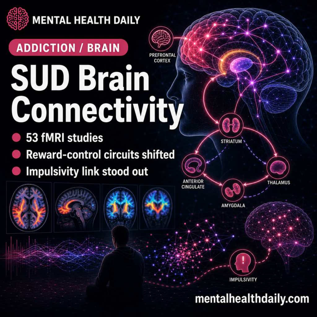
A 53-study resting-state functional magnetic resonance imaging (fMRI) meta-analysis found substance use disorder (SUD) linked to altered connectivity across prefrontal, striatal, thalamic, cingulate, and amygdala circuits. The most clinically relevant finding was an impulsivity correlation, not a diagnostic brain signature.
Research Highlights
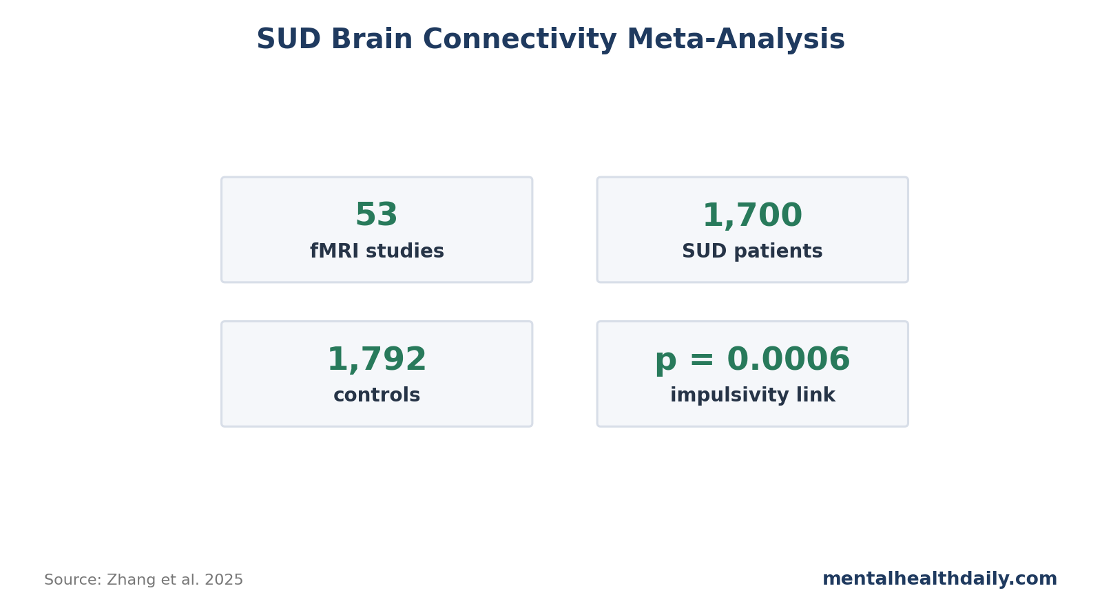
- Meta-analysis scale was meaningful: Zhang et al. pooled 53 whole-brain resting-state functional magnetic resonance imaging (rs-fMRI) studies, including 1,700 SUD patients and 1,792 healthy controls.1
- Reward-control circuitry was broadly altered: across 53 studies, anterior cingulate cortex (ACC), prefrontal cortex (PFC), striatum, thalamus, and amygdala seed analyses all showed abnormal connectivity patterns.
- Impulsivity had a circuit correlate: Barratt Impulsiveness Scale-11 (BIS-11) scores were negatively correlated with reduced resting-state connectivity between the striatum and median cingulate/paracingulate cortex, p = 0.0006.
- Family-wise error correction narrowed the map: after stricter family-wise error (FWE; multiple-comparison correction) testing, the 53-study map kept parts of the cortical-striatal-cortical pattern while some broader signals became less secure.
- Evidence strength is moderate but indirect: this meta-analysis maps group-level circuitry in 1,700 SUD patients and 1,792 controls; it cannot diagnose SUD, prove cause, or tell whether connectivity changes preceded drug exposure.
The analysis supports a circuit-level model of addiction neurobiology: SUD involves reward, control, salience, thalamic-relay, and affect-processing systems rather than one isolated addiction center.
53 Resting-State fMRI Studies Converged on Reward-Control Circuits
Resting-state fMRI measures synchronized blood-oxygen fluctuations while a person is not doing an explicit task. The method estimates functional connectivity: whether activity in 2 regions rises and falls together. It does not show direct wiring, and it does not show what a person is thinking.
Zhang et al. used Seed-based d Mapping, a coordinate-based meta-analytic method, to pool whole-brain seed-based rs-fMRI studies in SUD.1 The seed regions were selected because they sit in addiction-relevant reward and control circuitry:
- Anterior cingulate cortex: conflict monitoring, salience, emotional regulation, and impulse control.
- Prefrontal cortex: planning, inhibition, valuation, and top-down control.
- Striatum: reward learning, habit, motivation, and action selection.
- Thalamus: relay and gating of sensory, cognitive, and motivational signals.
- Amygdala: threat, emotional salience, and affective learning.
The broad result was dysfunction in cortical-striatal-thalamic-cortical circuitry. That phrase can sound abstract, but the logic is straightforward: addiction changes the loop between motivation, action, control, and emotional salience. Koob and Volkow described addiction as a neurocircuitry disorder involving binge/intoxication, withdrawal/negative affect, and preoccupation/anticipation stages.2 The 2025 meta-analysis adds a resting-state connectivity map to that model.
ACC and PFC Findings Fit Impaired Control and Salience Attribution
ACC pattern: the ACC showed increased connectivity with the inferior frontal gyrus, lentiform nucleus, and putamen. The right ACC/paracingulate cluster remained significant after FWE correction, with z = 4.042, p < 0.0001, and jackknife sensitivity in 15 of 16 datasets. Jackknife sensitivity means the result was retested while leaving out datasets one at a time.
PFC pattern: the PFC showed 2 directions at once: hyperconnectivity with the superior frontal gyrus and right striatum, plus hypoconnectivity with the bilateral inferior frontal gyrus. The right inferior frontal gyrus result remained significant after FWE correction, z = −4.208, p = 0.008.
That split fits the impaired response inhibition and salience attribution (iRISA) model. Goldstein and Volkow argued that addiction involves prefrontal dysfunction affecting self-control, salience attribution, awareness, and response inhibition.3 Zilverstand et al. later reviewed 105 task-based neuroimaging studies and found impairments across reward, habit, salience, executive, memory, and self-directed networks.4
Interpretation: the resting-state result complements task data. Even when participants are not performing an inhibition or cue-reactivity task, the background coupling of control and reward regions differs from controls.
Striatal Connectivity Linked the Map to Impulsivity
Striatum role: the striatum is often overcompressed into “reward center” language. In addiction, it is better understood as a hub for reward prediction, habit formation, action selection, and cue-driven motivation. In Zhang et al., striatal seeds showed hyperconnectivity with the superior frontal gyrus and hypoconnectivity with the median cingulate gyrus.
Impulsivity link: the clinically useful result came from 7 studies that included BIS-11 scores. Higher impulsivity was significantly associated with lower resting-state functional connectivity between the striatum and left median cingulate/paracingulate cortex, p = 0.0006. After FWE correction, the correlation remained significant.
That finding gives the connectivity map behavioral traction. It does not say impulsivity is caused by one broken connection. It says the group-level striatal-cingulate signal moved with a known clinical trait in SUD. Sutherland et al. argued earlier that resting-state connectivity may help capture the motivational, affective, and cognitive complexity of addiction better than isolated activation findings.5
The negative direction is important. Higher BIS-11 impulsivity tracked lower striatal-to-median-cingulate connectivity, which fits a weakened control-and-monitoring link rather than a simple “more reward activity” story. The result also narrows the article’s practical interpretation: the meta-analysis is most useful as a map of circuit traits that may affect relapse risk, cue reactivity, and treatment engagement, not as an argument that SUD can be reduced to reward-seeking alone.
Thalamus and Amygdala Results Extend the Model Beyond Reward
The thalamus showed reduced connectivity with the superior frontal gyrus, dorsal ACC, and caudate nucleus. The amygdala showed hypoconnectivity with the superior frontal gyrus and ACC, plus altered parahippocampal connectivity.
These results broaden the map in 2 ways:
- Thalamic gating: reduced thalamic-frontal and thalamic-caudate connectivity may affect how motivational and cognitive signals are routed through control loops.
- Affective salience: amygdala-frontal and amygdala-cingulate changes fit a model in which emotional cues, threat, stress, and drug cues are not cleanly separated.
Koob and Volkow’s neurocircuitry model gives that pattern clinical meaning. Withdrawal and negative affect are part of the addiction cycle. A person may continue substance use because reward, stress, dysphoria, cue reactivity, and impaired control interact.
Cue-memory context: the 2025 meta-analysis also found parahippocampal involvement across several seed regions. That does not make memory the dominant SUD mechanism, but it fits the clinical reality that drug cues are learned in places, routines, social contexts, withdrawal states, and stress states.
Circuit language becomes more useful when it connects reward-control findings to cue memory and affective context rather than treating addiction as one isolated dopamine loop. That framing also fits treatment planning better because relapse triggers are usually mixed, not anatomically pure.
Connectivity Findings Point to Treatment Targets, Not Brain-Scan Triage
Treatment-target logic: a circuit map can guide hypotheses about neuromodulation, cognitive training, cue-exposure work, relapse prevention, and medication mechanisms, but it should not be turned into brain-scan triage for individual patients. Mahoney et al. reviewed transcranial magnetic stimulation (TMS), deep brain stimulation, and related neuromodulation approaches for SUD and emphasized that stimulation targets need to be tied to substance type, clinical stage, and behavioral outcome.7
Invasive-treatment caution: Zhang et al. also cited emerging work on deep brain-machine interfaces for SUD, but the 2025 meta-analysis is not an argument for invasive treatment as a default.8 It is a reason to test whether specific circuit patterns predict who benefits from specific interventions.
A person with strong cue-reactivity, poor inhibitory control, and high impulsivity may need a different treatment sequence than a person whose main relapse driver is withdrawal distress, chronic pain, trauma reminders, or social exposure.
Clinical measurement: if resting-state connectivity eventually adds value, it will probably be as one layer next to craving scales, relapse history, sleep, pain, psychiatric comorbidity, medication exposure, and objective substance-use outcomes. A scan without behavior is too detached from the clinical problem.
Limitations of This Connectivity Meta-Analysis
Resting-state fMRI is correlational. Functional connectivity is a statistical association between signals. It cannot prove that one region drives another.
Substances were pooled. The included studies covered different substances, illness stages, abstinence durations, and scan protocols. A common SUD map is useful, but alcohol, opioids, stimulants, cannabis, and nicotine are not biologically identical exposures.
Coordinates lose detail. Coordinate-based meta-analysis uses reported peak locations rather than raw imaging data. It is practical, but it cannot harmonize preprocessing, medication status, withdrawal state, or scanner-level noise as well as pooled raw data could.
Group maps do not diagnose individuals. The study does not justify an fMRI test for addiction. It identifies group-level abnormalities that may guide mechanism research and intervention targets.
Questions About SUD Brain Connectivity
Does this prove addiction is a brain disease?
It supports a brain-circuit component of addiction, especially around reward-control loops. It does not reduce addiction to brain imaging alone. Social exposure, trauma, drug supply, psychiatric comorbidity, pain, housing, and treatment access still shape risk and recovery.
Can resting-state fMRI diagnose substance use disorder?
No. The findings are group-level differences. SUD remains a clinical diagnosis based on behavior, impairment, risk, craving, withdrawal, tolerance, and loss of control.
Why does the impulsivity correlation matter?
It links the imaging map to a measurable trait. A connectivity difference is more useful when it tracks behavior, symptom severity, relapse risk, treatment response, or cognitive performance.
What should future studies do?
Future work should separate substances, abstinence stages, sex, age, medication status, trauma exposure, and psychiatric comorbidity. Longitudinal studies are especially important because they can test whether connectivity differences predict relapse, recovery, or response to neuromodulation and behavioral treatment.
References
- Common Neural Patterns of Substance Use Disorder: A Seed-Based Resting-State Functional Connectivity Meta-Analysis. Zhang X et al. Translational Psychiatry. 2025;15:190. doi:10.1038/s41398-025-03396-2
- Neurobiology of Addiction: A Neurocircuitry Analysis. Koob GF, Volkow ND. Lancet Psychiatry. 2016;3:760-773. doi:10.1016/S2215-0366(16)00104-8
- Dysfunction of the Prefrontal Cortex in Addiction: Neuroimaging Findings and Clinical Implications. Goldstein RZ, Volkow ND. Nature Reviews Neuroscience. 2011;12:652-669. doi:10.1038/nrn3119
- Neuroimaging Impaired Response Inhibition and Salience Attribution in Human Drug Addiction: A Systematic Review. Zilverstand A et al. Neuron. 2018;98(5):886-903. doi:10.1016/j.neuron.2018.03.048
- Resting-State Functional Connectivity in Addiction: Lessons Learned and a Road Ahead. Sutherland MT et al. NeuroImage. 2012;62(4):2281-2295. doi:10.1016/j.neuroimage.2012.01.117
- Neurobiologic Advances From the Brain Disease Model of Addiction. Volkow ND et al. New England Journal of Medicine. 2016;374:363-371. doi:10.1056/NEJMra1511480
- Transcranial Magnetic Stimulation, Deep Brain Stimulation, and Other Forms of Neuromodulation for Substance Use Disorders: Review of Modalities and Implications for Treatment. Mahoney JJ III et al. Journal of the Neurological Sciences. 2020;418:117149. doi:10.1016/j.jns.2020.117149
- Harnessing the Sensing and Stimulation Function of Deep Brain-Machine Interfaces: A New Dawn for Overcoming Substance Use Disorders. Chen D et al. Translational Psychiatry. 2024;14:440. doi:10.1038/s41398-024-03156-8