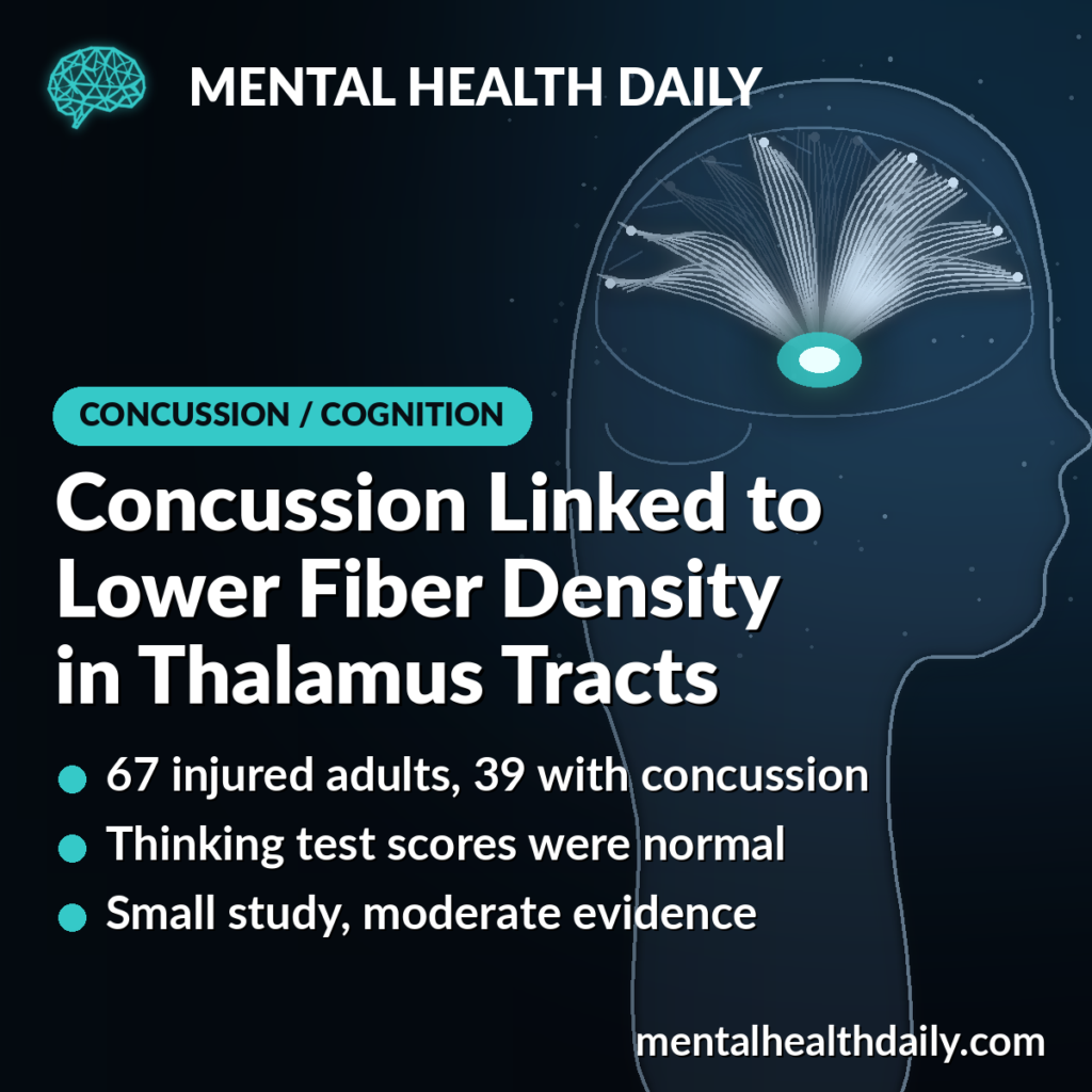
About 2 months after a concussion, adults had lower fiber density in a few white matter tracts linking the thalamus to the top of the brain, even though their thinking test scores looked normal. A 2026 study compared 39 people with mild traumatic brain injury to 28 people hurt in accidents without a head injury.1
Research Highlights
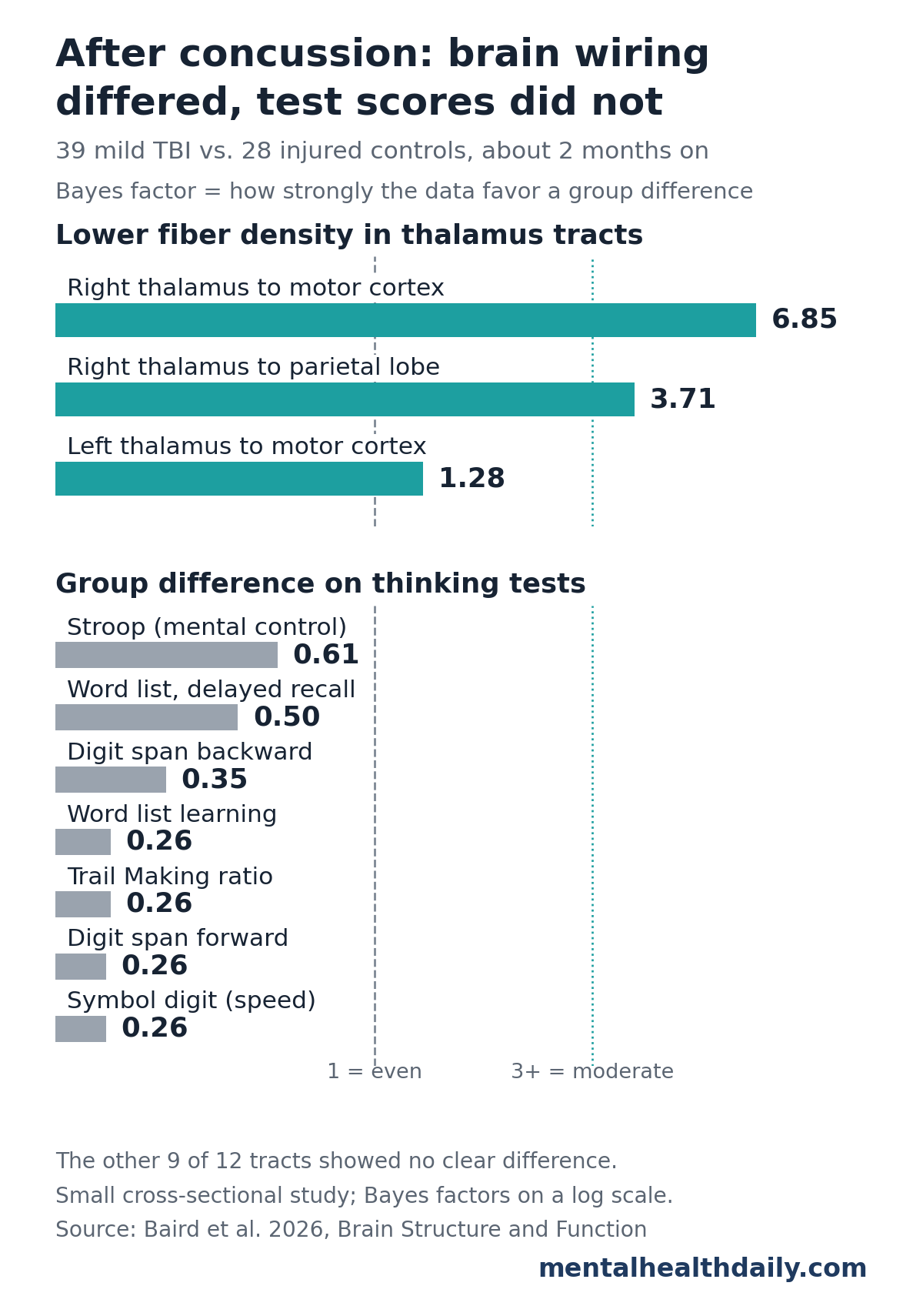
- Lower fiber density in 2 right-side tracts: moderate evidence for the tract from the thalamus to the right motor cortex (Bayes factor 6.85) and to the right parietal lobe (3.71). Evidence for the left motor tract was weak (1.28).1
- Most tracts showed no clear change: of 12 thalamus tracts, 5 favored no difference and 4 were inconclusive. A whole-brain scan analysis found no significant differences.1
- Thinking tests were similar: the concussion and injury-control groups scored alike on all 7 measures of attention, processing speed, memory and mental control.1
- Brain-test links differed by group: in exploratory models, higher fiber density in a left frontal thalamus tract went with slower processing speed after concussion but faster speed in controls.1
- Small study: 67 people scanned once, with no pre-injury scans. The results point to subtle tract changes, not a diagnostic test.1
How Fixel-Based Analysis Measures Thalamus Wiring
The thalamus is a pair of egg-shaped structures deep in the middle of the brain. It works as a relay hub, passing signals to and from nearly every part of the cortex, and it helps with attention, movement and processing speed.
Thalamocortical tracts are the bundles of nerve fibers (white matter) that connect the thalamus to the cortex. Because the thalamus sits in the center of the brain, its long tracts may be especially exposed to the twisting forces of a blow to the head.1
Most concussions leave no damage visible on a standard CT or MRI. To look for subtler changes, researchers use diffusion MRI, which tracks how water moves along nerve fibers. Older diffusion methods average every fiber direction inside each tiny cube of brain (a voxel), which blurs the picture where fibers cross.
Fixel-based analysis splits each voxel into its separate fiber bundles, called fixels, and measures each one on its own. Its main measure here was:
- Fiber density: an MRI estimate of how much axon (nerve fiber) material runs in a given direction. Lower fiber density may reflect a loss of axons.1
How the Melbourne Concussion Study Was Set Up
Maggie Baird et al. recruited adults admitted to 2 Melbourne, Australia, hospitals after an injury between 2017 and 2023.1
- Concussion group: 39 adults with mild traumatic brain injury, defined by a Glasgow Coma Scale score (a 15-point measure of alertness) of 13 to 15 plus brief confusion, memory loss or loss of consciousness. 67% scored a full 15.
- Control group: 28 adults hurt in similar accidents (car, motorbike, cycling, falls) with no blow to the head.
- Timing: one MRI and test session 6 to 12 weeks after injury, about 8 weeks on average.
- Ages: 18 to 60, average about 37 in both groups.
- Tracts: 6 thalamus tracts on each side of the brain, mapped with an automated tool, running to the prefrontal, premotor, motor, sensory, parietal and occipital cortex.
Using injured people as controls is a strength. Pain, fatigue and distress after an accident can all affect test scores, so this design helps isolate the effect of the head injury itself.
Fiber Density Was Lower in 2 Right-Side Thalamus Tracts
The researchers used Bayes factors to weigh the evidence. A Bayes factor compares how well the data fit “the groups differ” against “the groups do not differ.” A value of 1 means the data cannot tell them apart.
Above 3 counts as moderate evidence for a difference, and below 1/3 counts as evidence for no difference.1
After adjusting for age and sex, the concussion group had lower fiber density in:
- Right thalamus to motor cortex: Bayes factor 6.85 (moderate)
- Right thalamus to parietal lobe: Bayes factor 3.71 (moderate)
- Left thalamus to motor cortex: Bayes factor 1.28 (weak)
The other 9 tracts showed no clear difference. The motor cortex controls voluntary movement, and the parietal lobe helps combine sensory information and direct attention.
Whole-brain analysis: when the researchers tested every fixel across the brain, no differences survived correction for multiple comparisons. They suggest that concussion causes small changes spread along whole tracts, which show up when a tract is averaged but are too faint at any single point.1
Timing: the researchers propose that fiber density rises in the first days after a concussion from swelling, then falls if axons degenerate. By about 2 months, their results fit that later, lower phase.1
Thinking Test Scores Were Normal After Concussion
Both groups completed 7 measures from 5 standard tests of attention (digit span), processing speed (symbol digit), word learning and recall, mental control (Stroop) and task switching (Trail Making). None showed a group difference; Bayes factors ranged from 0.26 to 0.61.1
For example, the concussion group averaged 66.3 on the symbol digit speed test and the control group averaged 65.3. This fits the usual course after concussion, when most people’s test performance has recovered within about 3 months.3
Different brain-test links: the researchers then asked whether fiber density tracked test scores the same way in both groups. It did not always:1
- Processing speed: after concussion, higher fiber density in the left thalamus-to-premotor tract went with slower symbol digit scores (Bayes factor 10.4). In controls, the same tract went with faster scores (2.36).
- Attention: in controls, higher fiber density in several right-side tracts went with lower digit span scores. No such link appeared after concussion.
The study team called these results exploratory. Many models were run across 12 tracts and 7 tests, and the expected pattern in controls (more fiber density, better scores) mostly did not appear, possibly because of pain, fatigue and distress after their own injuries.1
Thalamus Changes After Concussion in Other Studies
The thalamus keeps turning up in concussion research:
- Older diffusion MRI: a meta-analysis of diffusion tensor imaging studies found the largest cluster of white matter change after mild brain injury centered on the left thalamus, extending into a thalamus tract.4
- Same method in athletes: fixel-based analysis in professional Australian football players found tract changes consistent with swelling within 12 days of injury that were largely gone in players scanned later.5
- Same cohort, different measure: in the same 67 people, overall thalamus connection strength was normal, but stronger connections went with better test scores in controls and worse scores after concussion.2
- Symptoms: in a study of 108 people with mild brain injury, early thalamus connectivity on functional MRI helped separate those who still had symptoms 6 months later.6
Together, these studies place the thalamus among the brain areas most affected by concussion. How tract changes relate to symptoms and recovery is still unclear.
Limitations of This Concussion Imaging Study
- Small sample: 67 people limits power, especially for the brain-test interaction models.
- One time point: with a single scan and no pre-injury baseline, the study cannot rule out differences that existed before the injury or show whether the changes fade or persist.
- Moderate, not strong, evidence: the 2 main Bayes factors were 3.7 and 6.8, and the whole-brain test was negative.
- Fiber density only: the tract method could not measure fiber cross-section, a related size measure.
- Specific group: hospitalized adults injured mostly in road accidents and falls; athletes, veterans, assault victims and people with repeated concussions were excluded.
What This Means After a Concussion
Normal thinking test scores 2 months after a concussion do not guarantee that the brain looks exactly as it did before. In this study, subtle wiring changes were still detectable in a few thalamus tracts, and the link between brain structure and test performance differed from that in injured controls.
These are group-level research findings. Fixel-based analysis is not a clinical test, and the study does not show that the changes cause symptoms or need treatment. Anyone with lingering headaches, fatigue, trouble concentrating or slowed thinking after a concussion should raise them with a doctor.
References
- Baird M, Seal ML, Beare R, Yang JY-M, Anderson JFI. Thalamocortical white matter microstructure and cognition following mild traumatic brain injury: a fixel-based analysis. Brain Structure and Function. 2026;231:142. doi:10.1007/s00429-026-03193-7
- Baird ME, Yang JY-M, Seal ML, Beare R, Anderson JFI. Thalamic structural connectivity and cognitive outcome in the subacute period following mild traumatic brain injury. Journal of Neurotrauma. 2026;43(11–12):882–894. doi:10.1177/08977151251407626
- Karr JE, Areshenkoff CN, Garcia-Barrera MA. The neuropsychological outcomes of concussion: a systematic review of meta-analyses on the cognitive sequelae of mild traumatic brain injury. Neuropsychology. 2014;28(3):321–336. doi:10.1037/neu0000037
- Aoki Y, Inokuchi R. A voxel-based meta-analysis of diffusion tensor imaging in mild traumatic brain injury. Neuroscience & Biobehavioral Reviews. 2016;66:119–126. doi:10.1016/j.neubiorev.2016.04.021
- Mito R, Parker DM, Abbott DF, Makdissi M, Pedersen M, Jackson GD. White matter abnormalities characterize the acute stage of sports-related mild traumatic brain injury. Brain Communications. 2022;4(4):fcac208. doi:10.1093/braincomms/fcac208
- Woodrow RE, Winzeck S, Luppi AI, et al. Acute thalamic connectivity precedes chronic post-concussive symptoms in mild traumatic brain injury. Brain. 2023;146(8):3484–3499. doi:10.1093/brain/awad056