A 2026 Nature Aging study involving 520 participants found that plasma protein-structure signatures classified healthy control, mild cognitive impairment, and Alzheimer’s disease groups with 83.44% three-way accuracy.1 The result is a serious biomarker signal, but it is not a clinic-ready replacement for established Alzheimer’s testing.
Research Highlights
- Classification was strong: The plasma structural-proteomics panel reached 83.44% accuracy across healthy control, mild cognitive impairment, and Alzheimer’s disease groups.1
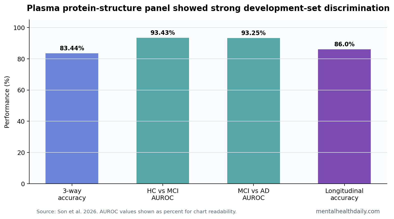
- Pairwise AUCs were high: Healthy control vs. MCI reached AUROC 0.9343, and MCI vs. Alzheimer’s disease reached AUROC 0.9325.1
- Longitudinal samples held up: Follow-up plasma samples were classified with 86.0% accuracy in the reported longitudinal subset.1
- Specific proteins drove the signal: Structural changes involving C1QA, clusterin, and ApoB peptides helped define the 3-protein panel.1
- Clinical translation remains separate: A 520-person biomarker-development study still needs external validation against p-tau, amyloid, imaging, and clinical outcomes.
Plasma structural proteomics means measuring protein accessibility and folding-state clues in blood under a chemical-labeling assay. In Alzheimer’s disease, that is a different question from ordinary concentration-based blood tests.
Mild cognitive impairment (MCI) is a measurable decline in cognition that is greater than expected for age but not severe enough to meet dementia criteria. The MCI boundary is clinically important because biomarker triage is most useful before irreversible functional decline is obvious.
83.44% Three-Way Accuracy Is the Headline Number
Son et al. used covalent protein profiling to label accessible peptide regions in plasma proteins, then trained machine-learning classifiers across healthy control, MCI, and Alzheimer’s disease groups. The final reported classifier reached 83.44% accuracy across the 3 groups.1
That number is stronger than a weak screening signal. A 3-way task is harder than a simple case-control comparison because MCI sits between normal aging and clinically apparent Alzheimer’s disease. The model also reported high pairwise discrimination: AUROC 0.9343 for healthy control vs. MCI and AUROC 0.9325 for MCI vs. Alzheimer’s disease.
AUROC means area under the receiver operating characteristic curve, a summary of how well a test separates groups across thresholds. A value near 0.5 is chance-level; values above 0.9 are usually considered strong in development datasets.
The important phrase is development dataset. Biomarker models often perform best in the dataset that generated them. Real-world memory clinics include vascular cognitive impairment, Lewy body disease, depression, sleep apnea, medication effects, traumatic brain injury, normal aging, and mixed pathology. A clinically useful blood test has to survive that messier population.
That does not weaken the result into noise. It sets the next threshold. A panel with AUROCs above 0.93 deserves replication because the signal is large enough to compete with established blood biomarker work. It still needs to show incremental value after age, APOE genotype, plasma p-tau217, amyloid-beta ratios, cognitive screening, and clinical history are already known.
C1QA, Clusterin, and ApoB Made the Panel Biologically Plausible
The panel centered on structural changes in C1QA, CLUS, and ApoB peptides. C1QA is part of complement biology, an immune pathway involved in tagging material for clearance. Clusterin, also called apolipoprotein J, is linked to lipid transport, amyloid handling, and inflammatory stress. ApoB is a major apolipoprotein in lipid-carrying particles.
The biological plausibility is useful because an Alzheimer’s blood classifier should not feel like a black-box lottery ticket. Complement, lipid transport, vascular risk, amyloid handling, and inflammation are all plausible routes into neurodegeneration. Still, plausibility does not validate a clinical test by itself.
The study also reported a broad accessibility gradient across 879 labeled peptides: average accessibility was 93.2% in healthy controls, 92.0% in MCI, and 91.1% in Alzheimer’s disease.1 That direction suggests a global structural-protein shift across the measured peptide set.
Why C1QA is interesting: complement biology sits near synapse pruning, immune activation, and clearance pathways. Alzheimer’s disease includes amyloid deposition, glial response, vascular change, and inflammatory signaling. A complement-linked plasma structure signal therefore has a plausible route into brain pathology, even if the blood measurement is indirect.
Why clusterin is interesting: clusterin has appeared repeatedly in Alzheimer’s genetics and biomarker discussions because it interacts with lipid handling and amyloid-related biology. A structural change in clusterin may tell a different story than a simple concentration shift.
Why ApoB is interesting: ApoB connects the panel to lipid-transport and vascular-risk biology. Dementia risk is partly neurodegenerative and partly vascular, and the same patient can have amyloid pathology, tau pathology, vascular injury, inflammation, and metabolic risk at the same time.
Protein Structure Adds a Different Layer Than p-Tau217
Alzheimer’s blood testing has been dominated by amyloid and tau markers. The ATN framework separates amyloid, tau, and neurodegeneration biology so a clinical syndrome is not confused with a single pathology label.2 Plasma p-tau217 has become one of the strongest blood candidates because it tracks Alzheimer’s tau pathology and often separates Alzheimer’s disease from non-Alzheimer conditions.3
The Son study asks a different question: can protein structure in plasma provide a parallel disease-stage signal? The answer in this dataset was yes, but the next comparison should be direct. A new protein-structure classifier is most useful if it adds information beyond p-tau217, amyloid-beta ratios, neurofilament light, APOE genotype, age, sex, and cognitive testing.
Plasma p-tau217 is the obvious benchmark because it is already close to clinical implementation in some settings.3 A protein-structure panel could still earn a role if it improves early MCI classification, identifies inflammatory or lipid-dominant subgroups, or flags people whose standard amyloid/tau markers are borderline.
Another possible role is staging rather than diagnosis. If structural accessibility shifts from healthy control to MCI to Alzheimer’s disease, repeated measures could eventually test whether the panel changes as symptoms progress. The current longitudinal subset was small but directionally encouraging, with 86.0% classification accuracy.
APOE Genotype Signals Need Careful Interpretation
The C1QA signal varied by APOE genotype, and APOE epsilon4/epsilon4 carriers showed the lowest accessibility. APOE epsilon4 is the strongest common genetic risk variant for late-onset Alzheimer’s disease, but it is not destiny. Many carriers never develop dementia, and many Alzheimer’s cases do not carry epsilon4.
That makes the genotype result interesting rather than determinative. A structural-protein panel might partly capture pathways connected to genetic risk, complement activation, or lipid biology. It cannot be treated as a genetic-risk verdict without prospective outcome data.
External Validation Is the Real Test
Evidence-strength note: this was a biomarker-development study. It can show that protein-structure signatures separated groups in the analyzed cohorts. It cannot show that the panel will maintain the same accuracy in primary care, memory clinics, mixed dementia, depression-related cognitive complaints, vascular cognitive impairment, Parkinson’s disease dementia, or diverse racial and socioeconomic populations.
Blood biomarkers also create a false-precision hazard. If a test is used in the wrong population, even high sensitivity and specificity can generate many false positives. The strongest clinical use would probably be as triage: identifying who needs amyloid/tau confirmation, specialist review, or longitudinal follow-up.
External validation should therefore report more than overall accuracy. It should report performance by age, sex, APOE genotype, race, kidney function, inflammatory disease, vascular disease, depression status, and medication exposure. Those factors can change plasma proteins and could make a structural-proteomics assay look cleaner in research cohorts than in routine care.
The test also needs calibration against outcomes that readers care about: conversion from MCI to dementia, rate of cognitive decline, future amyloid/tau positivity, and ability to distinguish Alzheimer’s disease from non-Alzheimer dementias. High AUCs are promising; longitudinal clinical discrimination is the harder target.
Another issue is workflow. A blood test can be excellent analytically and still be hard to use if the next step is unclear. A positive plasma protein-structure result should probably trigger confirmatory amyloid/tau testing or specialist review, not an immediate dementia label. A negative result should be interpreted in the context of symptoms, age, family history, medication burden, sleep, depression, and vascular risk.
That is where the study is most useful now. It expands the blood-biomarker conversation beyond amyloid and tau without replacing them. If protein-structure signatures replicate, they may become a complementary layer that explains inflammatory, lipid, or complement biology in patients whose ordinary Alzheimer’s markers tell only part of the story.
Comparator design should be strict. The next useful study would test the panel against p-tau217, amyloid-beta ratios, neurofilament light, APOE genotype, and ordinary clinical variables in the same people. A new assay earns clinical space only if it improves classification, staging, or prognosis beyond those easier-to-interpret signals.
Questions About Alzheimer’s Plasma Protein Structure Tests
Is this a new Alzheimer’s blood test?
It is a candidate blood biomarker panel. It is not yet a validated clinical replacement for established diagnostic workups.
Why does protein structure matter?
Protein folding and accessibility can change when inflammation, lipid transport, complement activation, or disease-related binding states change. Measuring structure may catch biology that ordinary concentration tests miss.
What would make the result stronger?
Independent validation, head-to-head comparison with plasma p-tau217 and amyloid markers, and prospective prediction of conversion from MCI to dementia would make the panel more clinically meaningful.
References
- Son J, et al. Structural signature of plasma proteins in Alzheimer’s disease. Nature Aging. 2026. doi:10.1038/s43587-026-01078-2
- Jack CR Jr, et al. A/T/N: an unbiased descriptive classification scheme for Alzheimer disease biomarkers. Lancet Neurology. 2016. doi:10.1212/wnl.0000000000002923
- Palmqvist S, et al. Discriminative accuracy of plasma phospho-tau217 for Alzheimer disease vs. other neurodegenerative disorders. JAMA. 2020. doi:10.1001/jama.2020.12134
- Nakamura A, et al. High performance plasma amyloid-beta biomarkers for Alzheimer’s disease. Nature. 2018. doi:10.1038/nature25456