A 2026 developing Human Connectome Project analysis of 438 full-term neonates found that the newborn cortex already had an adult-like folding hierarchy: gyri connected most strongly to other gyri, sulci connected most weakly to other sulci, and structure-function coupling shifted near 41 to 42 weeks postmenstrual age.1
Research Highlights
- Gyri were already high-connectivity hubs: across 438 neonates scanned from 38.14 to 44.71 weeks, gyro-gyral functional and structural connections were strongest, gyro-sulcal links were intermediate, and sulco-sulcal links were weakest.1
- The analysis used 64 cortical regions: researchers divided 32 neonatal atlas regions into gyral and sulcal regions of interest, then built 64 x 64 functional-connectivity and structural-connectivity matrices.1
- The hierarchy held across 7 weekly bins: from W38 through W44, whole-brain connectivity kept the same gyro-gyral > gyro-sulcal > sulco-sulcal ordering.1
- Coupling changed over days, not years: whole-brain structure-function coupling was positive for 56% of the 38-44 week window, with functional connectivity peaking near 42 weeks while structural connectivity kept increasing.1
- The limbic lobe was the outlier: reported coupling rates were 0.81 occipital, 0.59 temporal, 0.56 whole brain, 0.54 parietal, 0.52 frontal, and 0.13 limbic.1
Gyri are the raised folds of the cerebral cortex, while sulci are the grooves between those folds. Folding is not decorative anatomy: it changes wiring distance, local circuitry, blood-flow geometry, and how brain regions are packed into the skull.
Structure-function coupling means that physical wiring and synchronized activity change together. In MRI terms, structural connectivity usually comes from diffusion imaging of white-matter tracts, while functional connectivity comes from correlated blood-oxygen-level-dependent signals during resting-state functional MRI.
438 Newborn MRI Scans Showed Gyri-to-Gyri Links Were Strongest
Mao et al. analyzed 438 full-term neonates from the developing Human Connectome Project. All included infants were born after 38 weeks of gestation and scanned between 38.14 and 44.71 weeks postmenstrual age, giving the researchers a narrow but clinically important window around term-equivalent brain development.1
The study combined 3 MRI layers: T2-weighted structural imaging, diffusion-weighted imaging, and resting-state functional MRI. Structural MRI defined the cortical surface, diffusion MRI estimated white-matter fibers, and resting-state fMRI estimated how strongly regional activity rose and fell together.
Region construction: the neonatal atlas contained 32 cortical regions. Each region was split into a gyral and a sulcal component by cortical curvature, yielding 64 regions of interest. The researchers then compared 3 connection classes:
- Gyro-gyral: raised fold to raised fold.
- Gyro-sulcal: raised fold to nearby or distant groove.
- Sulco-sulcal: groove to groove.
The main result was simple and robust: both functional connectivity and structural connectivity followed the same ordering across the whole brain. Gyro-gyral connections were strongest, gyro-sulcal connections were intermediate, and sulco-sulcal connections were weakest. A 2-way analysis of functional connectivity found a folding-type main effect with p < 0.001 and a lobe main effect with p < 0.05.1
The Newborn Cortex Already Resembled the Adult Gyral-Hub Model
The finding matters because it pushes an adult brain-network principle back into the neonatal period. Deng et al. previously proposed an adult cortical model in which gyri act as global communication hubs and sulci act more like local processing units.2 Mao et al. found the same broad hierarchy before ordinary postnatal experience could plausibly explain it.
Adult-like hierarchy, active maturation: newborn gyri and sulci were already differentiated, but neonatal connectivity was still rapidly reorganizing. The study’s age window covered only 7 weekly bins, yet functional and structural trajectories changed enough for the structure-function relationship to flip direction in several regions.
The lobe-level pattern was not perfectly uniform. Temporal, frontal, and parietal lobes followed the global hierarchy cleanly. The occipital lobe kept the expected structural-connectivity order, but its functional-connectivity pattern was slightly different: gyro-gyral links remained strongest, while sulco-sulcal functional connectivity was slightly higher than gyro-sulcal functional connectivity. Limbic connectivity was also less cleanly separated at individual weekly time points.1
That regional unevenness is useful. A newborn brain is not a scaled-down adult brain. Primary sensory networks, association networks, limbic circuitry, and cortical folding patterns mature on different schedules, so a single global average can hide the regions where development is most asynchronous.
Structure-Function Coupling Shifted Around 41 to 42 Weeks
The most informative part of the paper was the changing relationship between physical wiring and synchronized activity across the term-age window.
Mao et al. fit developmental curves from 38 to 44 weeks. Across the whole brain, functional connectivity increased from 38 weeks, peaked near 42 weeks, and then declined. Structural connectivity increased more steadily across the period. During the early part of the window, the 2 measures moved together. After roughly 42 weeks, they separated: structural connectivity kept rising while functional connectivity no longer followed the same path.1
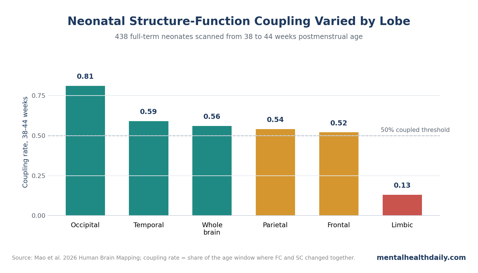
Coupling rates exposed regional timing: the occipital lobe was coupled for 81% of the window, while the limbic lobe was coupled for only 13%. Temporal, frontal, and parietal lobes sat near the middle at 59%, 52%, and 54%, respectively. Whole-brain coupling was 56%.
At the regional level, 11 of 32 cortical regions were classified as more decoupled than coupled, 8 were roughly balanced, and 13 were more coupled than decoupled. That split is a warning against reading neonatal brain development as one synchronized wave. Some regions are building structural wiring and functional synchrony in parallel; others are already moving through a mismatch phase.
Why the 41-Week Shift Fits Other Neonatal Connectome Work
Several adjacent imaging studies make the timing plausible. Batalle et al. reviewed neonatal imaging evidence and emphasized that the newborn period is an active developmental window; diffusion and functional imaging can capture early substrates of later cognitive and motor outcomes, especially in infants with perinatal risk.3
Franca et al. added a dynamic-connectivity layer. In a 2024 neonatal study, term and preterm infants differed in dynamic functional-connectivity organization, and early connectivity features related to neurodevelopmental outcomes in childhood.4 Mao et al. did not test later outcomes, but their 41- to 42-week coupling shift lands in the same conceptual space: the neonatal connectome is already changing on a timescale that could matter for later developmental trajectories.
Dear et al. pushed the bridge from normal development to psychiatry further by showing that healthy cortical gene-expression architecture overlaps with imaging, transcriptomic, and genetic signals in autism and schizophrenia.5 A neonatal gyral-sulcal map is therefore best read as normative developmental anatomy, not diagnosis. Normative cortical architecture is part of the same biological terrain where later neurodevelopmental and psychiatric risk can become visible.
This Is a Normative Map, Not a Newborn Mental-Health Screen
The strongest interpretation is anatomical and developmental. Gyri appear to function as higher-connectivity hubs by term-equivalent age, and the structure-function relationship changes rapidly across the 38-44 week window. That is already enough.
The weaker interpretation would be to treat this as an early diagnostic biomarker for autism, schizophrenia, depression, or cognitive delay. The study did not do that. It did not follow infants into childhood, compare diagnostic groups, or test predictive accuracy. It also used a relatively coarse 32-region atlas, and gyral-sulcal classification depended on curvature thresholds even though sensitivity checks across 0.1, 0.15, and 0.20 were consistent.1
Evidence-strength note: this was a large normative imaging analysis, not a clinical prediction study. It can support claims about term-age cortical organization. It cannot support claims about individual prognosis, psychiatric diagnosis, or treatment selection.
Clinical bridge: the valuable next step is longitudinal follow-up that links neonatal coupling patterns with later developmental outcomes.
That follow-up should separate ordinary term-age variation from prematurity, hypoxia, infection, and neonatal intensive-care exposures that can reshape early networks.
One paper-internal caution is worth flagging. The results section reported a limbic coupling rate of 0.13, meaning predominantly decoupled development, while a later discussion sentence described the limbic lobe as coupled through most of the period. The numeric result is the safer read: limbic structure-function timing looked atypical relative to the rest of the cortex, and future work should clarify that trajectory.
Questions About Neonatal Brain MRI and Gyri
Does this mean newborn gyri are already mature?
No. Gyri already showed stronger structural and functional links than sulci, but maturation was still active. Functional connectivity peaked and then declined near 42 weeks while structural connectivity kept increasing, which is the opposite of a static mature system.
Could this help predict autism or schizophrenia risk?
Not yet. Cortical folding and connectivity are relevant to neurodevelopmental disorders, but this study analyzed full-term neonates as a normative cohort. Prediction would require longitudinal follow-up, diagnostic outcomes, and independent validation.
Why compare gyri and sulci instead of ordinary brain regions?
Gyri and sulci are basic units of cortical folding. Comparing them asks whether brain-network architecture follows the geometry of the cortical sheet as well as named anatomical regions such as frontal or temporal cortex.
What would make the finding clinically stronger?
A stronger clinical test would link neonatal gyro-sulcal connectivity to later cognition, language, motor development, autism traits, or psychiatric symptoms. It would also need finer parcellation, preterm and high-risk cohorts, and replication across scanners.
References
- Mao W, He Z, Jin X, Hamouda E, Ou X, Kendrick KM, et al. Structuro-functional differentiation and coupling of gyri and sulci in the neonatal cortex. Human Brain Mapping. 2026;47:e70524. doi:10.1002/hbm.70524
- Deng F, Jiang X, Zhu D, et al. A functional model of cortical gyri and sulci. Brain Structure & Function. 2014;219:1473-1491. doi:10.1007/s00429-013-0581-z
- Batalle D, Edwards AD, O’Muircheartaigh J. Annual Research Review: not just a small adult brain: understanding later neurodevelopment through imaging the neonatal brain. Journal of Child Psychology and Psychiatry. 2018;59(4):350-371. doi:10.1111/jcpp.12838
- Franca LGS, Ciarrusta J, Gale-Grant O, et al. Neonatal brain dynamic functional connectivity in term and preterm infants and its association with early childhood neurodevelopment. Nature Communications. 2024;15:16. doi:10.1038/s41467-023-44050-z
- Dear R, Wagstyl K, Seidlitz J, et al. Cortical gene expression architecture links healthy neurodevelopment to the imaging, transcriptomics and genetics of autism and schizophrenia. Nature Neuroscience. 2024;27:1075-1086. doi:10.1038/s41593-024-01624-4
- Dierker DL, Feczko E, Pruett JR Jr, et al. Analysis of cortical shape in children with simplex autism. Cerebral Cortex. 2015;25(4):1042-1051. doi:10.1093/cercor/bht294