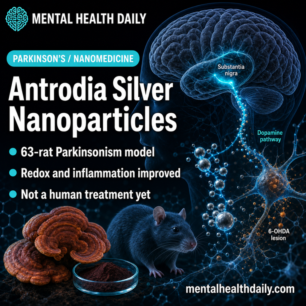
A 2026 Parkinsonism study involving 63 male rats found that Antrodia cinnamomea plus citrate-stabilized silver nanoparticles reduced 6-OHDA motor, oxidative-stress, inflammatory, α-synuclein, and apoptosis markers, while dopamine and acetylcholine recovery remained nonsignificant. The useful claim is a multitarget preclinical signal, not a Parkinson’s disease treatment recommendation.
Research Highlights
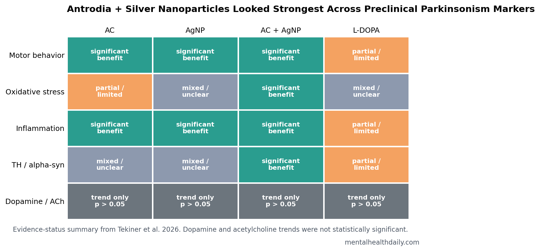
- Combination treatment looked strongest: Tekiner et al. tested 63 male rats across 9 groups and found the AC + AgNP arm reduced several 6-OHDA injury signals more consistently than either ingredient alone.1
- Motor and redox markers improved: apomorphine rotations differed among lesioned groups at p < 0.05, and AC + AgNP restored GSH and SOD antioxidant markers vs. 6-OHDA at p < 0.05.1
- Inflammation fell sharply: TNF-α and IL-1β were lower in the AC, AgNP, and AC + AgNP lesioned groups vs. 6-OHDA, with several comparisons at p < 0.001.1
- Neurotransmitters stayed uncertain: dopamine and acetylcholine showed partial recovery trends, but LC-MS/MS group differences did not reach significance (p > 0.05).1
- Translation is early: the evidence comes from SH-SY5Y cells and a short-term 6-OHDA male-rat model, not from human Parkinson’s patients or long-term nanoparticle safety data.1
Antrodia cinnamomea is a medicinal fungus native to Taiwan that contains triterpenoids, polysaccharides, and phenolic compounds with antioxidant and immunomodulatory activity. Silver nanoparticles are tiny silver-based particles used experimentally as drug-delivery and bioactivity platforms; citrate coating helps stabilize them, but dose, size, coating, and tissue distribution can change safety.
6-OHDA means 6-hydroxydopamine, a toxin that selectively injures catecholamine neurons and is commonly used to create Parkinsonism-like dopaminergic damage in rodents. It is useful for testing mechanisms around oxidative stress and dopamine-neuron injury, but it does not reproduce the slow, multifactorial course of human Parkinson’s disease.
63 Male Rats Tested AC, AgNPs, and the Combined Formulation
Tekiner et al. used 2 linked models: differentiated SH-SY5Y cells exposed to 6-OHDA and 63 adult male Sprague-Dawley rats assigned to 9 groups of 7 rats each.1 The in vivo experiment included nonlesioned control/treatment groups and lesioned groups receiving L-DOPA, Antrodia cinnamomea (AC), citrate-stabilized silver nanoparticles (AgNPs), or the AC + AgNP combination.
Intervention setup: rats received AC at 100 mg/kg, AgNPs at 1 mg/kg, or both for 14 days. On the final treatment day, the researchers injected 6-OHDA unilaterally into the substantia nigra, the midbrain region where dopamine-producing neurons degenerate in Parkinson’s disease.
Outcome spread: the study did not rely on one marker. It measured cell viability, apomorphine rotations, locomotor activity, cylinder forelimb asymmetry, brain malondialdehyde (MDA), reduced glutathione (GSH), superoxide dismutase (SOD), serum TNF-α and IL-1β, dopamine and acetylcholine by LC-MS/MS, histology, tyrosine hydroxylase, α-synuclein, agmatinase, Bcl-2, Caspase-3, and PI3K.
Why the design helps: the 9-arm layout separated background treatment effects from lesion-specific effects. Nonlesioned AC, AgNP, and AC + AgNP groups made it possible to see whether the materials changed behavior or biology without 6-OHDA injury, while lesioned treatment groups tested whether those same materials shifted the Parkinsonism-like injury pattern.
L-DOPA acted as a familiar comparator rather than a perfect benchmark, because short toxin-lesion models can respond differently from chronic human disease. That design makes the multitarget signal more informative than a simple before-after experiment, but it still leaves dose durability, sex effects, tissue accumulation, and long-term nanoparticle safety unanswered.
AC + AgNP Improved Motor, Redox, and Inflammation Readouts
In the cell model, 6-OHDA reduced SH-SY5Y viability compared with control cells (p < 0.001). AC + AgNP improved viability at p < 0.05, especially around 25-50 μM in the authors’ discussion, while AgNP treatment alone was associated with lower cell viability in some conditions.1
In rats, the 6-OHDA group showed the expected motor injury: increased apomorphine-induced rotations, lower locomotor activity, longer rest time, and forelimb-use asymmetry. AC + 6-OHDA, AgNP + 6-OHDA, and AC + AgNP + 6-OHDA reduced rotations vs. 6-OHDA, and rotations differed among lesioned groups at p < 0.05.
The redox and cytokine pattern was more consistent than the neurotransmitter pattern:
- Lipid peroxidation: MDA, a marker of lipid oxidative damage, rose with 6-OHDA and fell in multiple treatment groups.
- Antioxidant defense: GSH and SOD were reduced by 6-OHDA; AC + AgNP increased both vs. 6-OHDA at p < 0.05.
- Inflammation: TNF-α and IL-1β were elevated by 6-OHDA and reduced by AC, AgNP, and AC + AgNP comparisons, with key cytokine comparisons at p < 0.001.
Dopamine and Acetylcholine Recovery Was Only Trend-Level
The most important calibration point is the LC-MS/MS neurotransmitter result. LC-MS/MS means liquid chromatography-tandem mass spectrometry, an analytic method used here to quantify dopamine and acetylcholine in substantia nigra tissue.
Dopamine and acetylcholine were lower in the 6-OHDA group, and treated groups showed partial recovery trends. The combined AC + AgNP arm preserved a dopamine-increase trend, but group differences did not reach statistical significance (p > 0.05). That means the study supports a broad injury-marker signal more strongly than a confirmed neurotransmitter-restoration claim.
Practical interpretation: if a summary says the formulation restored dopamine, that is too strong. A tighter read is that AC + AgNP improved several upstream and tissue-level markers while neurotransmitter recovery remained statistically unconfirmed.
TH, Alpha-Synuclein, PI3K, and Caspase-3 Pointed to Cell-Survival Biology
Tyrosine hydroxylase (TH) is the rate-limiting enzyme for dopamine synthesis and is often used as a marker of dopaminergic neuron integrity. TH immunoreactivity was reduced by 6-OHDA and increased significantly in the L-DOPA + 6-OHDA and AC + AgNP + 6-OHDA groups vs. 6-OHDA; AC alone and AgNP alone did not reach significance for TH in the lesioned groups.1
Α-synuclein is the protein that accumulates in Lewy pathology in Parkinson’s disease. 6-OHDA increased α-synuclein immunoreactivity (p < 0.001), while AC + AgNP reduced it vs. 6-OHDA (p < 0.05). The result is not evidence of clearing human Lewy bodies, but it fits the broader protein-stress pattern in the toxin model.
The Western blot results also leaned toward survival-pathway modulation. PI3K is part of a cell-survival signaling pathway, Bcl-2 is an anti-apoptotic protein, and Caspase-3 is an executioner enzyme in apoptosis. 6-OHDA reduced PI3K, Bcl-2, agmatinase, and TH while increasing Caspase-3; treatment comparisons moved those markers away from the injury pattern.
Adjacent Nanoparticle and Antrodia Studies Make the Signal Less Isolated
Kara et al. tested astaxanthin-loaded silver nanoparticles in a related 6-OHDA Parkinsonism model and reported benefit through endoplasmic-reticulum stress and PI3K/Akt/mTOR signaling.2 Yeni et al. tested L-DOPA-modified zinc oxide nanoparticles in 6-OHDA rats, placing Tekiner et al. inside a growing nanocarrier literature rather than a one-off mushroom-extract experiment.3
The Antrodia side also has a nearby model. Lanza et al. reported that Antrodia camphorata extract affected Nrf2-mediated neuropathological changes in a Parkinson’s mouse model.4 Nrf2 is a transcription factor that helps cells turn on antioxidant-defense genes, so that paper lines up with the oxidative-stress emphasis in the Tekiner study.
Zhao et al. separately reported neuroprotective effects of biogenic silver nanoparticles in Parkinson’s disease in vitro and in vivo.5 The shared theme is not that all nanoparticles are safe or effective. It is that several preclinical Parkinsonism papers are testing whether nanocarriers can concentrate antioxidant, anti-inflammatory, or dopaminergic-support strategies in vulnerable neural tissue.
The 6-OHDA Model Is Useful, but It Is Not Human Parkinson’s Disease
Neurotoxin models are designed to create a controlled lesion. Bove and Perier described why these models remain useful for studying dopaminergic injury while also warning that they simplify the biology of Parkinson’s disease.6 Human Parkinson’s disease develops over years, involves prodromal non-motor symptoms, variable genetics, aging biology, gut-brain and immune factors, and a clinical course that no short toxin experiment fully recapitulates.
Several limits keep the Tekiner result in the preclinical lane:
- Male-only animals: 63 rats were all male, so sex-dependent effects were not tested.
- Short exposure window: treatment lasted 14 days, not months or years.
- Nanoparticle safety: silver nanoparticle effects vary by dose, size, coating, tissue accumulation, and duration; long-term toxicology remains central.
- Model specificity: 6-OHDA produces rapid dopaminergic injury rather than slow human disease progression.
- Neurotransmitter uncertainty: dopamine and acetylcholine recovery did not pass the p < 0.05 threshold.
The best next experiments would compare dose levels, particle sizes, coatings, female and male animals, longer follow-up, behavioral durability, pharmacokinetics, tissue distribution, and progressive Parkinson’s models. Human use would require a separate safety and efficacy program, not extrapolation from this rat study.
Questions About Antrodia Silver Nanoparticles and Parkinsonism
Does this study show that Antrodia supplements treat Parkinson’s disease?
No. The study tested a specific AC-loaded citrate-stabilized silver nanoparticle formulation in cells and male rats. It did not test over-the-counter Antrodia products in people with Parkinson’s disease.
Why combine Antrodia with silver nanoparticles?
The rationale is delivery plus multitarget biology. Antrodia compounds may affect oxidative stress and inflammation, while citrate-stabilized AgNPs may alter delivery and bioactivity. That combination still needs long-term safety testing.
Which result is most clinically tempting but easiest to overstate?
Dopamine. The treated groups showed partial recovery trends, but the LC-MS/MS differences were nonsignificant at p > 0.05. The stronger evidence sits in redox, cytokine, histology, TH, α-synuclein, and apoptosis markers.
Could this become a Parkinson’s drug?
Only after a long translational path. The formulation would need reproducible chemistry, toxicology, dose-finding, pharmacokinetics, progressive animal-model data, and eventually human trials. The current paper is a preclinical signal.
References
- Tekiner D, Gedikli S, Gelen V, Bayram C, Kara A. Modulation of Oxidative Stress and Apoptosis by Antrodia cinnamomea-Loaded Citrate-Stabilized Silver Nanoparticles in Experimental Parkinsonism. Molecular Neurobiology. 2026;63:576. doi:10.1007/s12035-026-05853-5
- Kara H, Tekiner D, Ustundag H, et al. Astaxanthin-loaded silver nanoparticles mitigate 6-OHDA-induced Parkinson’s via ER stress and PI3K/Akt/mTOR signaling. Molecular Neurobiology. 2025;63(1):156. doi:10.1007/s12035-025-05279-5
- Yeni Y, Genc S, Ertugrul MS, et al. Neuroprotective effects of L-Dopa-modified zinc oxide nanoparticles on the rat model of 6-OHDA-induced Parkinson’s disease. Scientific Reports. 2024;14:19077. doi:10.1038/s41598-024-69324-4
- Lanza M, Cucinotta L, Casili G, et al. The transcription factor Nrf2 mediates the effects of Antrodia camphorata extract on neuropathological changes in a mouse model of Parkinson’s disease. International Journal of Molecular Sciences. 2023;24(11):9250. doi:10.3390/ijms24119250
- Zhao S, Zhang J, Zhang J. Neuroprotective effects of biogenic silver nanoparticles in Parkinson’s disease: in vitro and in vivo study. International Journal of Biological Macromolecules. 2024;1:138236. doi:10.1016/j.ijbiomac.2024.138236
- Bove J, Perier C. Neurotoxin-based models of Parkinson’s disease. Neuroscience. 2012;211:51-76. doi:10.1016/j.neuroscience.2011.10.057