A 2026 mouse study found that Theiler’s murine encephalomyelitis virus (TMEV) injected into the substantia nigra produced persistent Parkinson’s-like motor signals through 20 weeks, including apomorphine-induced rotations at every measured post-injection timepoint.1 The finding makes TMEV a useful viral-neuroinflammation model of dopamine-neuron injury, but it does not show that Theiler’s virus causes human Parkinson’s disease.
Research Highlights
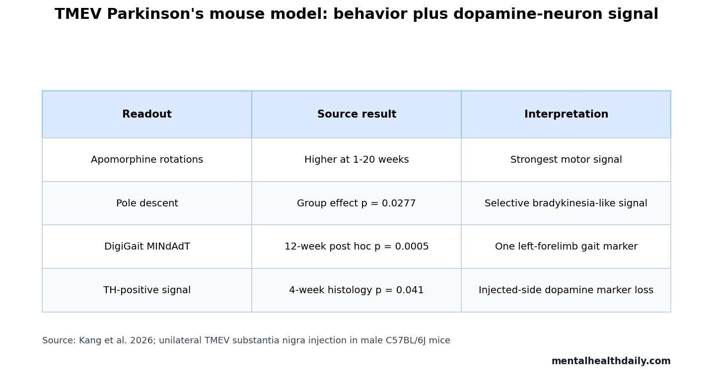
- 20-week lesion behavior persisted: TMEV-injected mice showed more apomorphine-induced contralateral rotations than controls at 1, 4, 8, 12, 16, and 20 weeks.
- Rotation statistics were strong: the apomorphine rotation analysis found a TMEV group effect of F(1,25) = 17.03, p = 0.0004, plus a group-by-time interaction of F(6,150) = 6.993, p < 0.0001.
- Pole-test impairment was narrower: descent time showed a TMEV group effect of F(1,25) = 5.462, p = 0.0277, while turn time was significant only at selected weeks.
- Gait data added one motor marker: among 48 DigiGait parameters, left-forelimb MINdAdT differed by TMEV group, with the 12-week post hoc comparison at p = 0.0005.
- Dopamine-neuron marker loss was visible: tyrosine-hydroxylase signal was lower on the injected side at 4 weeks in n = 5 TMEV mice vs. n = 3 controls, p = 0.041.
Theiler’s murine encephalomyelitis virus is a naturally occurring mouse virus often used to study virus-triggered neurological disease. In this study, researchers used it as an infectious trigger for a Parkinson’s disease model because prior work had already shown that TMEV can infect tyrosine-hydroxylase-positive neurons in the substantia nigra.2
Tyrosine hydroxylase is the rate-limiting enzyme for dopamine production and is commonly used as a marker for dopaminergic neurons. Substantia nigra pars compacta means the dopamine-rich midbrain region that degenerates in Parkinson’s disease and sends dopamine signals into the striatum, a movement-control hub.
TMEV Produced Persistent Apomorphine Rotations Through 20 Weeks
Kang et al. injected roughly 1400 plaque-forming units of TMEV in 0.5 microliters into the right substantia nigra pars compacta of adult male C57BL/6J mice. Control mice received the same surgery with culture medium instead of virus.
The main behavioral experiment included n = 13 TMEV mice and n = 14 control mice. Apomorphine-induced rotation was the clearest signal. Apomorphine is a dopamine receptor agonist; in a unilateral dopamine lesion model, it can make the dopamine-depleted side behave as if its receptors have become hypersensitive, producing rotations away from the lesioned side.
TMEV injection increased contralateral rotations at every measured post-injection point: 1 week (p = 0.0255), 4 weeks (p = 0.0027), 8 weeks (p = 0.0222), 12 weeks (p = 0.0024), 16 weeks (p = 0.0027), and 20 weeks (p = 0.0006). Baseline rotations did not differ before surgery.
That time-course pattern is the strongest reason to treat the model as more than a transient infection effect. The virus was no longer detected by the 4-week histology point, yet the motor readout persisted into the 20-week endpoint.
Pole-Test and Gait Signals Were More Selective
The pole test measures how long a mouse takes to turn downward and descend a vertical pole, a simple assay often used for bradykinesia-like motor slowing in Parkinson’s models. In the TMEV study, descent time carried the cleaner group-level result.
For time-to-descend, TMEV injection had a significant group effect: F(1,25) = 5.462, p = 0.0277. Post hoc testing separated TMEV from control mice at 4 weeks (p = 0.0100), 16 weeks (p = 0.0080), and 20 weeks (p = 0.0315).
Time-to-turn was less stable. The group effect trended in the expected direction, F(1,25) = 3.431, p = 0.0758, while the group-by-time interaction reached p = 0.0452. Week-specific tests were significant at 4 weeks and 16 weeks, with a 20-week trend.
DigiGait is a treadmill system that quantifies paw placement and movement across gait cycles. Out of 48 measured gait parameters, the left-forelimb maximal rate of paw-area change during propulsion, labeled MINdAdT, was lower in TMEV mice. The group effect was F(1,20) = 4.592, p = 0.0446, with the 12-week post hoc difference at p = 0.0005.
Tyrosine-Hydroxylase Loss Anchored the Motor Behavior to Dopamine Neurons
The behavioral signal would be much weaker if it were not paired with dopamine-neuron evidence. At 1 week, one mouse was used for qualitative confirmation that the surgical approach put TMEV into tyrosine-hydroxylase-positive neurons in the substantia nigra.
At 4 weeks, researchers compared the injected and uninjected sides of the substantia nigra. TMEV mice had a higher uninjected-to-injected tyrosine-hydroxylase intensity ratio than controls, p = 0.041, consistent with lower dopaminergic signal on the injected side. The histology comparison was small, n = 5 TMEV mice and n = 3 controls, but it matched the direction of the rotational behavior.
Evidence-strength note: this was an animal-only mechanistic model. It can support the claim that a mouse-specific neurotropic virus can create a Parkinson’s-like unilateral dopamine-lesion phenotype under experimental injection conditions. It cannot support a claim that TMEV, or viral infection generally, causes ordinary human Parkinson’s disease.
Injection route caveat: the model used direct substantia nigra delivery, not nasal, gut, blood-borne, or naturally acquired infection. That makes it useful for testing how infected dopamine-region tissue behaves, but it bypasses the real-world exposure steps that would have to occur before a human viral-causation argument became credible.
The 1997 TMEV Study Supplied the Missing Precursor
Oliver et al. reported in 1997 that Theiler’s virus could specifically infect tyrosine-hydroxylase-positive neurons in the mouse substantia nigra and cause degeneration, with limited spread to other brain regions.2 That paper supplied the target-cell specificity.
Kang et al. added the behavioral validation that the older paper did not fully establish. The 2026 study asked whether TMEV infection of substantia nigra dopaminergic neurons also produces motor readouts similar to unilateral toxin-lesion models.
The behavioral time course is the useful addition. Apomorphine rotations persisted from week 1 through week 20, which gives the model a chronic window rather than a brief acute-injury readout. For drug or immune-modulation experiments, that longer window matters because candidate treatments can be tested after the lesion phenotype has stabilized.
Model value: TMEV gives researchers a way to study how viral infection, dopamine-neuron loss, and neuroinflammation can intersect in the same experimental system. That is different from saying it reproduces the full slow, bilateral, alpha-synuclein-rich biology of human Parkinson’s disease.
Viral Parkinsonism Is Plausible, but Human Causation Remains Unsettled
Viral parkinsonism has a real literature. Jang et al. reviewed examples in which viral encephalopathies were followed by transient or persistent parkinsonism, including influenza and other neurotropic infections.3 Leta et al. later emphasized the same calibration problem: post-infectious parkinsonism, acute parkinsonian syndromes, and idiopathic Parkinson’s disease risk are related questions, but they are not the same question.4
Why the model still helps: a pathogen can damage basal ganglia circuits in a case report or animal model without being the usual cause of Parkinson’s disease in humans. A viral model can test mechanisms that toxin models often miss: glial activation, innate immune signaling, viral clearance, and delayed neuronal injury.
Preclinical comparison: toxin models such as 6-OHDA and MPTP remain useful because they reliably injure nigrostriatal dopamine neurons, but they create injury through chemical toxicity rather than infection-centered inflammation.5 TMEV adds a different route into the same dopamine-lesion phenotype.
What the TMEV Parkinson’s Model Still Needs
The most important missing comparison is direct benchmarking against established lesion models. Kang et al. did not run TMEV side by side with 6-OHDA or MPTP in the same experiment, so model severity, timing, and assay sensitivity still need direct calibration.
- Natural infection route: stereotaxic substantia nigra injection is experimentally precise, but it is not how ordinary viral infections reach the human nervous system.
- Sex and strain limits: the behavioral experiment used male C57BL/6J mice only, so female vulnerability, resistant vs. susceptible strains, and genetic-background effects remain open.
- Inflammation mechanism: the study did not measure glial states, cytokine timing, apoptosis markers, or striatal dopamine content, all of which would clarify how infection produced sustained injury.
- Human translation: TMEV is a mouse pathogen with no known human disease role, so the model is better read as a viral-neuroinflammation tool than as a candidate human cause.
Gerhauser et al. reviewed how TMEV can model several neurological disease phenotypes, including demyelination and seizures, depending on viral strain, host genetics, immune response, and tissue context.6 The Parkinson’s model fits that broader pattern: TMEV is not one disease in a bottle; it is a neurotropic trigger whose outcome depends on where, how, and in which host it is studied.
Questions About Viral Parkinson’s Mouse Models
Did the 2026 TMEV study show that viral infections cause Parkinson’s disease?
No. It showed that direct injection of a mouse-specific virus into the substantia nigra can produce persistent dopamine-lesion behavior in mice. Human Parkinson’s disease causation remains a separate question.
Why did apomorphine rotations matter so much?
Apomorphine rotations are a classic readout of unilateral nigrostriatal dopamine damage. In this study, rotations stayed higher in TMEV mice through 20 weeks, making the behavioral signal more durable than a short acute-sickness effect.
Does this model prove neuroinflammation is enough to create Parkinson’s disease?
No. It supports viral neuroinflammation as one experimentally useful route into dopaminergic injury. It does not reproduce every hallmark of human Parkinson’s disease, and it still needs glial, apoptosis, dopamine-content, and alpha-synuclein pathology work.
References
- Kang TW, Srinivasan R, Brinkmeyer-Langford C, Welsh CJ. Theiler’s murine encephalomyelitis virus as the infectious agent for a virally induced mouse model of Parkinson’s disease. Brain, Behavior, & Immunity – Health. 2026;54:101230. https://doi.org/10.1016/j.bbih.2026.101230
- Oliver KR, Brennan P, Fazakerley JK. Specific infection and destruction of dopaminergic neurons in the substantia nigra by Theiler’s virus. Journal of Virology. 1997;71(8):6179-6182. https://doi.org/10.1128/jvi.71.8.6179-6182.1997
- Jang H, Boltz DA, Webster RG, Smeyne RJ. Viral parkinsonism. Biochimica et Biophysica Acta. 2009;1792(7):714-721. https://doi.org/10.1016/j.bbadis.2008.08.001
- Leta V, Urso D, Batzu L, et al. Viruses, parkinsonism and Parkinson’s disease: the past, present and future. Journal of Neural Transmission. 2022;129(9):1119-1132. https://doi.org/10.1007/s00702-022-02536-y
- Duty S, Jenner P. Animal models of Parkinson’s disease: a source of novel treatments and clues to the cause of the disease. British Journal of Pharmacology. 2011;164(4):1357-1391. https://doi.org/10.1111/j.1476-5381.2011.01426.x
- Gerhauser I, Hansmann F, Ciurkiewicz M, Loscher W, Beineke A. Facets of Theiler’s murine encephalomyelitis virus-induced diseases: an update. International Journal of Molecular Sciences. 2019;20(2):448. https://doi.org/10.3390/ijms20020448