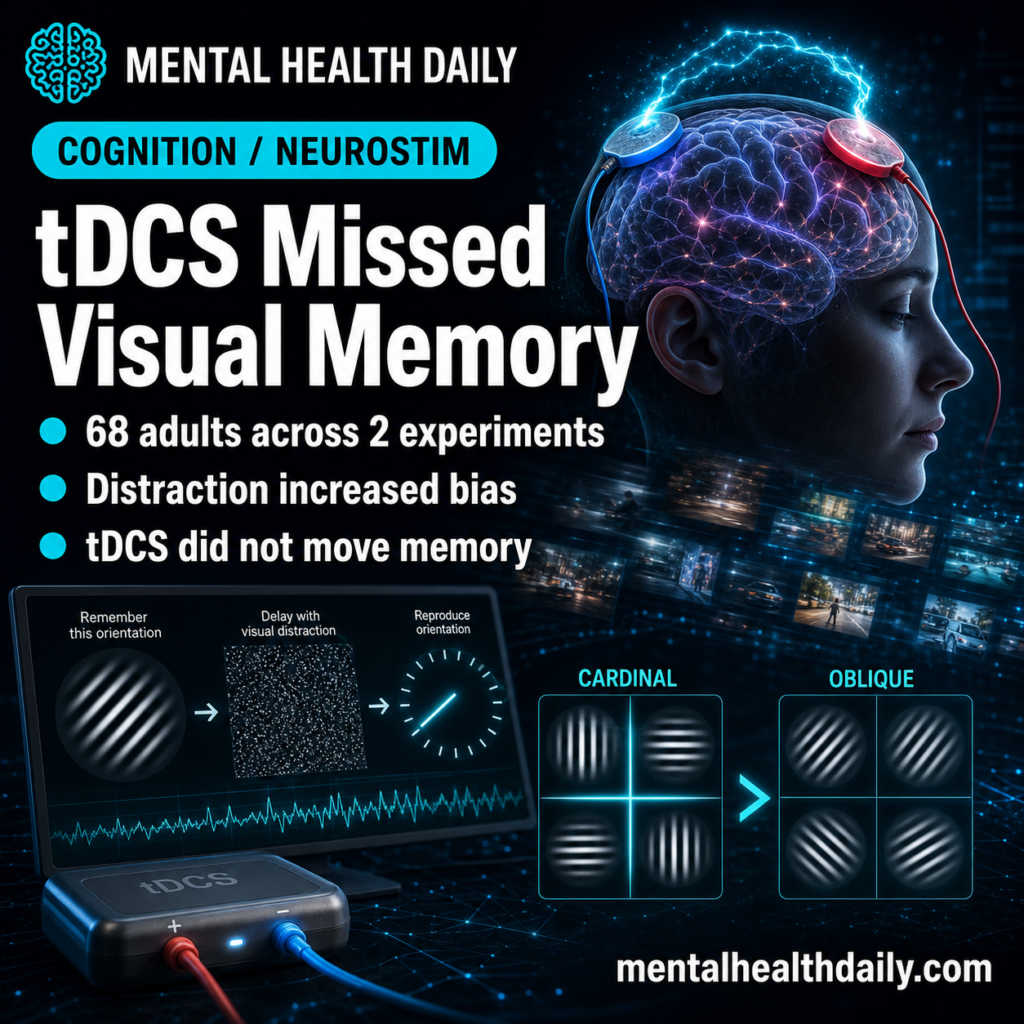
A 2026 two-experiment study found no visual-working-memory benefit from parietal or occipital transcranial direct current stimulation, while distraction unexpectedly made the cardinal-over-oblique orientation advantage stronger than it was without distraction: the pooled distraction-by-angle interaction had BFinc = 1021.671.1
Research Highlights
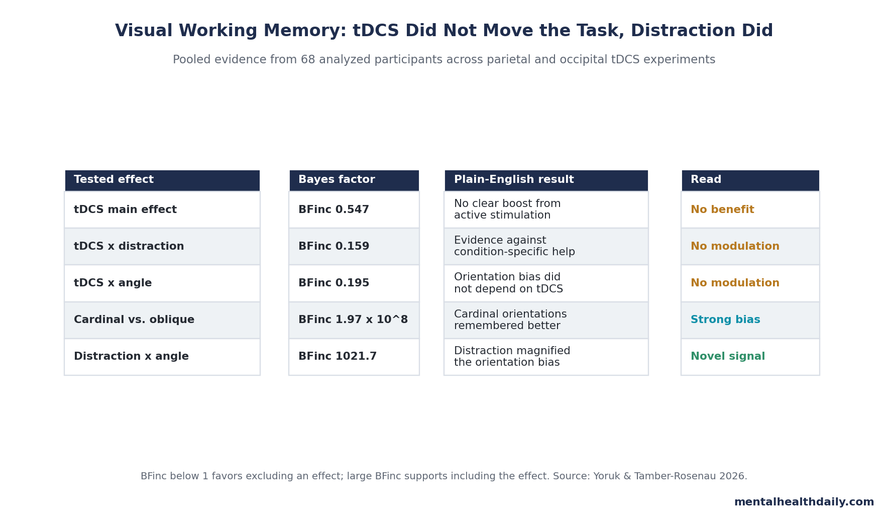
- Distraction strengthened the bias: pooled data from 68 analyzed adults showed a strong distractor-by-angle interaction, with BFinc = 1021.671 and a larger cardinal-over-oblique advantage during distractor-present blocks than during distractor-absent blocks.1
- tDCS did not improve memory: active stimulation had weak evidence against a main effect in the pooled analysis, BFinc = 0.547, after parietal and occipital experiments with 35 and 33 analyzed participants.1
- Target-specific rescue failed: tDCS did not clearly interact with distraction, BFinc = 0.159, or with cardinal vs. oblique orientation, BFinc = 0.195, so the stimulation did not selectively protect the difficult conditions.1
- Cardinal orientations dominated: across both experiments, cardinal angles were remembered better than oblique angles, with BFinc = 1.968 x 10⁸ for the angle effect.1
- Strategy may explain the paradox: distractors may have pushed memory toward categorical labels such as horizontal or vertical, which can preserve task performance while losing fine-grained oblique detail.1,4
Visual working memory is the short-term holding system that lets a person keep visual information online after the image is gone. In this study, participants briefly saw oriented Gabor patches, held one orientation in mind for 6 seconds, and then judged whether a probe was rotated clockwise or counterclockwise from the remembered target.
Transcranial direct current stimulation (tDCS) applies a weak electrical current through scalp electrodes to change cortical excitability. The practical question was whether a 20-minute, 2 mA anodal tDCS session over the posterior parietal cortex or occipital cortex could improve fine visual-memory judgments, especially when distracting pictures appeared during the delay.
68 Adults Tested Parietal and Occipital tDCS
Yörük and Tamber-Rosenau ran 2 closely matched within-subject experiments. In Experiment 1, 35 adults were analyzed after 2 exclusions for chance-level performance. Each participant completed an active anodal tDCS session over right posterior parietal cortex and a sham session.
Experiment 2 repeated the task with occipital stimulation over Oz, analyzing 33 adults after 4 exclusions. In both experiments, participants completed blocks with visual distractors and blocks without distractors, and each trial required a fine orientation judgment from memory.
Why the design was informative: the posterior parietal cortex is often linked to attentional control and visual-memory storage, while the occipital cortex contains early visual areas that code orientation. If either region was the bottleneck, active stimulation should have moved performance, reduced the oblique effect, or protected memory during distraction.
tDCS Did Not Improve Visual Working Memory
The stimulation result was the negative half of the paper’s answer. In the pooled analysis, active tDCS vs. sham had BFinc = 0.547, which did not support adding a tDCS main effect to the model. The tDCS-by-distraction interaction was even less supportive, BFinc = 0.159, and the tDCS-by-angle interaction was BFinc = 0.195.1
Bayes factor here means evidence for including a tested effect in the statistical model. A value below 1 leans against the effect; very small values provide stronger evidence that the tested term is unnecessary.
That pattern undercuts a simple “stimulate the memory region, improve the memory” account. Posterior parietal stimulation did not rescue distraction-heavy trials. Occipital stimulation did not sharpen orientation precision. Neither montage weakened the cardinal-over-oblique gap.
Distraction Increased the Oblique Effect Instead of Reducing It
Oblique effect means people perceive and remember horizontal and vertical orientations more accurately than diagonal orientations. The effect is familiar in vision research because neurons in early visual cortex tend to represent cardinal orientations more strongly than oblique ones.5,6
The stronger result was distraction’s direction: it made the oblique effect larger. In pooled data, the cardinal-over-oblique advantage was stronger in distractor-present trials, with Cohen’s d = 0.992 and BFinc = 7.931 x 10⁸, than in distractor-absent trials, where Cohen’s d = 0.551 and BFinc = 800.231.1
That direction was opposite the hypothesis. The researchers expected distractors to push memory away from early visual cortex and toward parietal representations, which should have reduced the oblique effect if the bias mainly came from early visual cortex. Instead, the behavioral bias grew.
Plain-English read: distraction preserved enough visual memory for the task while pulling the remembered orientation toward easy cardinal categories.
Prior fMRI Work Predicted a Cleaner Parietal Shift
Bettencourt and Xu decoded visual short-term memory under distraction and found that meaningful distractors disrupted occipital representations while parietal representations persisted, without obvious behavioral impairment.2 That result helped motivate the idea that parietal cortex can protect memory when new visual input competes with early visual cortex.
Rademaker et al. complicated that picture by showing coexisting sensory and mnemonic information in visual cortex, with meaningful-picture distractors reducing occipital mnemonic information and producing some behavioral cost.3 Hallenbeck et al. then showed that distraction can alter working-memory codes without fully erasing recoverable information, and that trial-to-trial memory errors can remain linked to visual-cortex representations.4
The 2026 tDCS study sits directly inside that disagreement, but its contribution is causal and behavioral rather than decoding-based. If parietal or occipital excitability alone controlled the shift, active stimulation should have changed the result. It did not.
Null tDCS Fits a Mixed Neuromodulation Literature
The tDCS result should not be stretched into a claim that noninvasive stimulation never affects working memory. It does fit a messy literature in which some studies reported benefits and others found nothing.
Tseng et al. reported that right posterior parietal tDCS improved change-detection performance especially in low-performing individuals.7 Wang et al. later reported selective enhancement of visual short-term-memory capacity after posterior parietal stimulation.8 Other work has been less encouraging: Jiang et al. found that tDCS over posterior parietal cortex or dorsolateral prefrontal cortex did not improve visual working-memory capacity.9
Important calibration: Yörük and Tamber-Rosenau tested representational precision, not how many objects people could store. A null result on fine orientation discrimination does not automatically cancel capacity findings from different tasks.
It does raise the bar for easy enhancement claims. The 2026 experiments used within-subject active vs. sham sessions, standard 2 mA and 20-minute stimulation parameters, and larger samples than many older tDCS working-memory studies. The task was sensitive enough to detect the oblique effect and its modulation by distraction, so the null stimulation result cannot be dismissed as a completely blunt behavioral measure.
The Best Explanation Is a Flexible Memory Code
Sensory recruitment is the view that visual working memory partly reuses early visual cortex, the same kind of neural machinery that initially codes visual input. Frontoparietal accounts put more weight on frontal and parietal areas that hold task-relevant information in a more abstract control network. Distributed coding treats both as plausible, with the brain shifting memory format depending on task demands.10,11
The new data favor the distributed view more than a single-region story. Stimulation of one cortical target did not move performance, which could mean the task was supported by parallel codes robust enough to absorb a coarse excitability manipulation.
Distraction’s effect on the oblique bias points to a more specific mechanism:
- Sensory-code recovery: visual cortex representations may be disrupted during distraction but recovered when the probe appears, leaving cardinal-tuned visual cortex to shape the final comparison.
- Categorical compression: participants may rely more on labels such as horizontal or vertical when distractors are predictable, improving usable task performance while losing diagonal precision.
- Multiple-code buffering: parietal and occipital systems may both help preserve the remembered item, so changing one region with standard tDCS is too crude to shift behavior.
Clinical and Cognitive-Enhancement Claims Should Stay Narrow
Evidence strength: the study is useful for cognitive neuroscience, not clinical treatment. It tested single-session tDCS in young adults with no reported neurological or psychiatric diagnosis. It did not test ADHD, dementia, depression-related cognition, traumatic brain injury, or repeated stimulation.
That limit cuts both ways. The null result should make consumer-style “brain stimulation improves working memory” claims look weaker for this task. It should not be used to rule out carefully targeted neuromodulation in clinical populations, multi-session protocols, individualized montages, or stimulation methods that target oscillatory timing rather than tonic excitability.
The more durable reader-facing point is about memory format. Visual working memory behaves less like a tiny photograph in the head and more like an active code that can trade fine detail for a usable, biased representation under distraction.
Questions About tDCS, Distraction, and Visual Working Memory
Did tDCS improve visual working memory in this study?
No. Neither right posterior parietal nor occipital anodal tDCS improved the orientation-memory task compared with sham stimulation.
What was the most surprising result?
Distraction made the oblique effect stronger. Cardinal orientations were remembered better than oblique orientations in general, and the gap increased when visual distractors appeared during the memory delay.
Does that mean distraction improved memory?
No. The main effect of distractor presence was not supported. The sharper result is that distraction changed the pattern of errors: memory became more biased toward cardinal orientations.
Why does the oblique effect matter?
The oblique effect is a clue about representational format. If diagonal orientations suffer more than horizontal or vertical orientations, the memory trace may still be shaped by sensory visual-cortex coding or by categorical compression toward cardinal labels.
Should this change how people think about consumer tDCS?
It should increase skepticism toward broad cognitive-enhancement claims. A sensitive within-subject study found real behavioral structure in the task but no stimulation benefit from 2 common cortical targets.
References
- The oblique effect in visual working memory is enhanced by distraction, regardless of tDCS manipulations. Yörük H, Tamber-Rosenau BJ. Cognitive, Affective, & Behavioral Neuroscience. 2026. doi:10.3758/s13415-026-01443-z
- Decoding the content of visual short-term memory under distraction in occipital and parietal areas. Bettencourt KC, Xu Y. Nature Neuroscience. 2016;19:150-157. doi:10.1038/nn.4174
- Coexisting representations of sensory and mnemonic information in human visual cortex. Rademaker RL et al. Nature Neuroscience. 2019;22:1336-1344. doi:10.1038/s41593-019-0428-x
- Working memory representations in visual cortex mediate distraction effects. Hallenbeck GE et al. Nature Communications. 2021;12:4714. doi:10.1038/s41467-021-24973-1
- An oblique effect in human primary visual cortex. Furmanski CS, Engel SA. Nature Neuroscience. 2000;3:535-536. doi:10.1038/75702
- Oblique effect: A neural basis in the visual cortex. Li B et al. Journal of Neurophysiology. 2003;90:204-217. doi:10.1152/jn.00954.2002
- Unleashing potential: Transcranial direct current stimulation over the right posterior parietal cortex improves change detection in low-performing individuals. Tseng P et al. Journal of Neuroscience. 2012;32:10554-10561. doi:10.1523/JNEUROSCI.0362-12.2012
- Electrical stimulation over human posterior parietal cortex selectively enhances the capacity of visual short-term memory. Wang S et al. Journal of Neuroscience. 2019;39:528-536. doi:10.1523/JNEUROSCI.1959-18.2018
- TDCS over PPC or DLPFC does not improve visual working memory capacity. Jiang S et al. Communications Psychology. 2024;2:20. doi:10.1038/s44271-024-00067-8
- The distributed nature of working memory. Christophel TB et al. Trends in Cognitive Sciences. 2017;21:111-124. doi:10.1016/j.tics.2016.12.007
- Reframing the debate: The distributed systems view of working memory. Lorenc ES, Sreenivasan KK. Visual Cognition. 2021;29:416-424. doi:10.1080/13506285.2021.1899091