
In rats with toxin-damaged cerebellums, 4 weeks of the blood-pressure drug losartan reduced oxidative damage, inflammation and tissue injury. A nanoparticle version did better on several tissue and staining measures, but not on gene activity.1
Research Highlights
- A small rat experiment: 24 male rats in 4 groups of 6. Three groups received carbon tetrachloride (a toxic industrial solvent) for 6 weeks; 2 of those then got oral losartan, either regular or as nanoparticles, for 4 weeks.1
- Both forms reduced the damage: compared with untreated toxin-exposed rats, treated rats had less oxidative stress, more Purkinje cells, better myelin staining and less activation of support cells.1
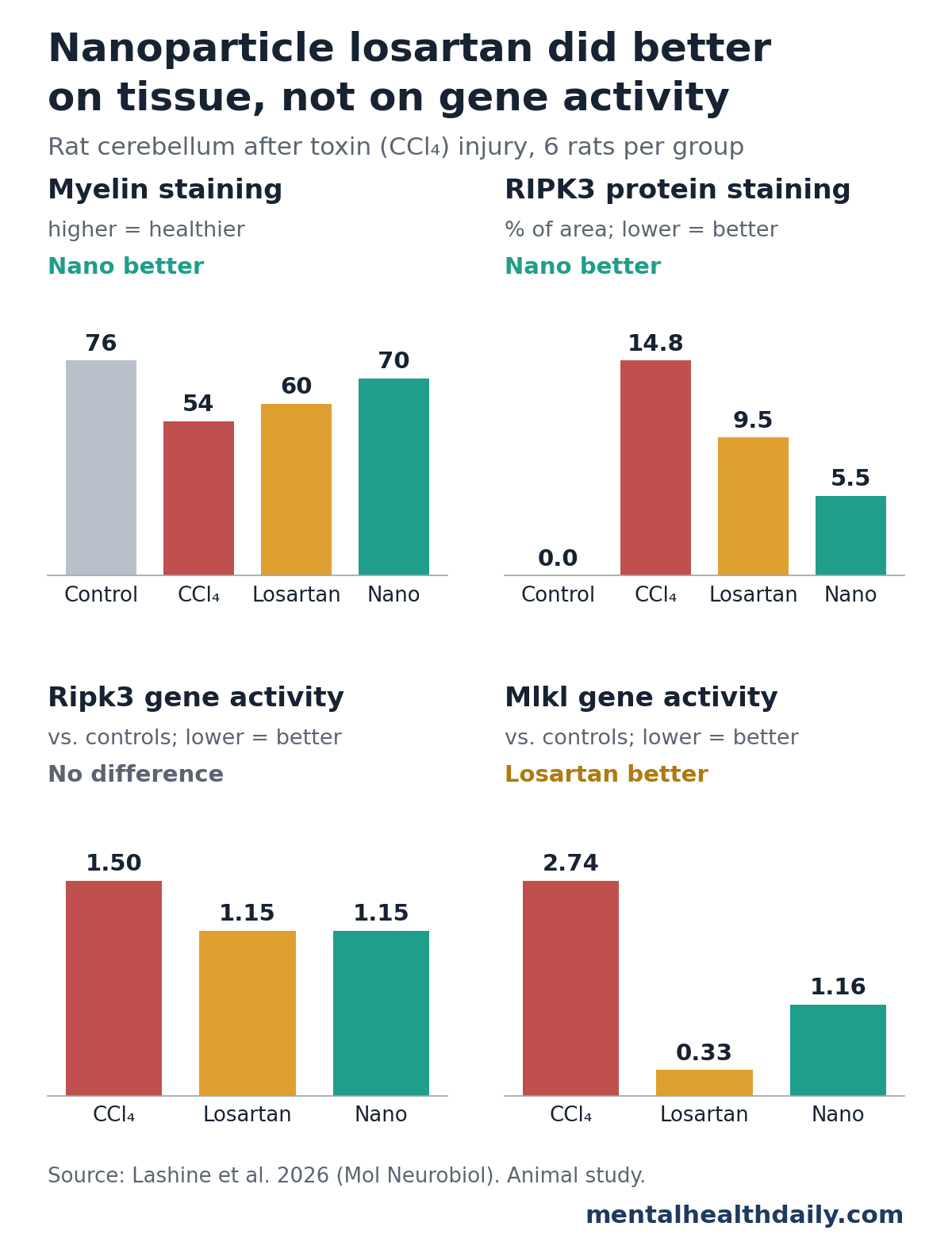
- Nanoparticles did better on most tissue measures: myelin staining was 69.5 with nanoparticles vs. 60.5 with regular losartan, and RIPK3-stained area was 5.5% vs. 9.5%.1
- No nanoparticle advantage in gene activity: Tnf, Ripk1 and Ripk3 gene activity fell about equally with both forms, and Mlkl fell further with regular losartan (0.33 vs. 1.16).1
- Necroptosis markers moved, but the pathway was not proven: the team did not measure the activated (phosphorylated) forms of the proteins that carry out necroptosis, and did not test movement or behavior.1
Carbon Tetrachloride, Losartan and Necroptosis Explained
A few terms first:
- Carbon tetrachloride (CCl4) is a solvent once used in dry cleaning and fire extinguishers. It dissolves in fat, passes easily into the brain and generates free radicals that damage cell membranes. Labs use it as a standard way to injure organs in rodents.12
- Losartan is a widely prescribed blood-pressure drug. It blocks the AT1 receptor for angiotensin II, a hormone that narrows blood vessels and also promotes inflammation and oxidative stress in tissues, including the brain.
- Necroptosis is a programmed form of cell death that ends with the cell bursting open and spilling inflammatory contents. It runs through 3 proteins in sequence: RIPK1 and RIPK3 join to form a complex, which switches on MLKL, and MLKL punches holes in the cell membrane.1
Caspase-8 acts as a brake on that sequence. When it is active, it cuts RIPK1 and RIPK3 apart and steers cells away from necroptosis.1
The cerebellum is the fist-sized structure at the back of the brain that coordinates movement and balance. Its large Purkinje cells are the main output neurons, and they are sensitive to toxins and oxidative stress.
How the Rat Experiment Was Set Up
Nermeen Lashine et al. at Mansoura University in Egypt randomly assigned 24 adult male Sprague-Dawley rats to 4 groups of 6:1
- Control: olive-oil injections twice a week for 6 weeks, then 4 weeks without treatment.
- CCl4: CCl4 injections twice a week for 6 weeks, then 4 weeks without treatment.
- Losartan: the same 6 weeks of CCl4, then regular losartan by mouth at 10 mg/kg a day for 4 weeks.
- Nanoparticle losartan: the same 6 weeks of CCl4, then the same daily dose given as losartan nanoparticles for 4 weeks.
Treatment started only after the injury phase ended, so the experiment tested whether losartan could help a cerebellum that was already damaged.1
The nanoparticles were pure losartan ground in a ball mill into tiny crystals, with no added carriers or coatings. Smaller particles have more surface area, which can change how a drug dissolves and is absorbed. The team confirmed their shape under an electron microscope but did not measure particle size distribution, drug levels in the blood or brain, or how the crystals behaved in the body.1
At week 10, researchers measured cerebellar tissue in several ways:
- Oxidative stress: malondialdehyde (MDA, a product of damaged fats) and 3 antioxidant defenses: glutathione, catalase and superoxide dismutase.
- Gene activity: messenger RNA levels for Tnf (an inflammatory signal), Ripk1, Ripk3 and Mlkl.
- Tissue structure: Purkinje cell counts, thickness of the granular layer and myelin staining.
- Protein staining: the share of tissue area stained for RIPK1, RIPK3 and caspase-8, plus GFAP (a marker of activated astrocytes) and Iba1 (a marker of microglia, the brain’s immune cells).
Investigators scoring the tissue did not know which group each slide came from.1
Carbon Tetrachloride Damaged the Rat Cerebellum
Compared with control rats, rats left untreated after CCl4 showed broad damage:1
- Fewer Purkinje cells: 5.0 per microscope field vs. 11.9 in controls.
- Thinner granular layer: 188 vs. 236 micrometers.
- Weaker myelin staining: 54.4 vs. 75.7.
- More oxidative damage: higher MDA and lower glutathione, catalase and superoxide dismutase.
- Necroptosis-related markers up: Mlkl gene activity rose to 2.74 relative units, RIPK3 protein covered 14.8% of the stained area (none was detected in controls), and caspase-8 staining fell from 7.2% to 0.4%.
- Activated support cells: GFAP staining rose from 7.5% to 20.8% and Iba1 from 6.6% to 16.9%.
Those findings are consistent with earlier rat work showing that even a single dose of CCl4 raises lipid damage and drains antioxidant enzymes across brain regions.2
Both Forms of Losartan Reduced Cerebellar Damage in Rats
Compared with untreated CCl4 rats, both treated groups improved on every type of measure: oxidative stress, gene activity, tissue structure and protein staining.1
- Purkinje cells: 7.6 per field with regular losartan and 10.0 with nanoparticles, vs. 5.0 untreated. The 2 forms did not differ significantly.
- Granular layer: 225 and 235 micrometers, both close to the control value of 236.
- Astrocyte and microglia activation: GFAP fell to 13.5% and 11.2%, and Iba1 to 11.6% and 8.5%.
- Caspase-8: rose back to 2.6% and 3.9%, still below the control level.
Most measures improved without returning fully to normal. RIPK3 staining, for example, stayed above the control level in both treated groups.1
Nanoparticle Losartan Did Better on Some Measures, Not All
Where nanoparticles came out ahead: compared with regular losartan, the nanoparticle group had significantly better results on:1
- Oxidative stress: lower MDA, and larger improvements in the antioxidant measures.
- Myelin staining: 69.5 vs. 60.5.
- RIPK1 staining: 1.2% vs. 3.2%, with the nanoparticle group no different from controls.
- RIPK3 staining: 5.5% vs. 9.5%.
- Caspase-8, GFAP and Iba1: all closer to control levels.
Where they did not: gene activity followed a different pattern. Tnf, Ripk1 and Ripk3 messenger RNA fell to similar levels with both forms; Ripk3, for instance, was 1.15 in each. Mlkl, the gene for the protein that actually ruptures the cell, fell further with regular losartan: 0.33 vs. 1.16.1
The researchers presented the nanoparticle advantage as an observation only. Without data on how much drug reached the brain, the study cannot say whether the crystals were absorbed better, acted differently or simply varied by chance in groups of 6.1
Necroptosis Markers Changed, but the Pathway Was Not Tested Directly
Higher RIPK1, RIPK3 and Mlkl levels show that the machinery for necroptosis was more abundant. They do not show it was switched on. Necroptosis is activated when RIPK3 and MLKL are phosphorylated (tagged with phosphate groups), and the team did not measure those activated forms.1
The researchers state this plainly: the results show an association with necroptosis-related changes, not proven activation or blockade of the pathway. The improvements could also follow from less oxidative stress and less tissue injury overall.1
Why necroptosis draws interest in brain disease: postmortem brains from people with Alzheimer’s disease show activated necroptosis that tracks with disease stage and lower cognitive scores, and blocking necroptosis reduced neuron loss in a mouse model of the disease.3
Earlier Research on Losartan and the Brain
According to the researchers, this is the first test of losartan against chemical injury to the cerebellum. Other animal studies point in a similar direction:
- Alzheimer’s mice: losartan at 10 mg/kg reduced amyloid plaques and inflammatory signals including TNF-alpha, increased new neuron formation and improved memory and learning compared with vehicle-treated mice.4
- Aged, inflamed mice: 4 weeks of losartan was linked to lower levels of nitrotyrosine (an oxidative stress marker) and tau protein in the frontal cortex.5
In people: a large cohort of mostly male US veterans found that those taking angiotensin receptor blockers, the drug class losartan belongs to, developed dementia at lower rates than those on other heart drugs (hazard ratio 0.76, or about 24% lower) or the ACE inhibitor lisinopril (0.81).6 That study was observational, so it cannot separate the drugs’ effects from differences between the people who took them, and it has no bearing on toxin injury.
Limitations of This Losartan Nanoparticle Rat Study
This is an early animal experiment. It can show that losartan changed tissue and marker measures after toxin injury in rats; it cannot show benefit for people or for brain function.1
- Small and narrow: 6 male rats per group, one toxin, one dose.
- No function measured: no tests of balance, coordination or behavior, which are what cerebellar damage actually affects.
- No pre-treatment baseline: no rats were examined at week 6, so the results show less damage than untreated rats at week 10, not reversal of measured injury.
- Semi-quantitative staining: protein results come from stained area, not direct protein measurement such as Western blots.
- Missing controls: no healthy rats given losartan alone and no sham-milled comparison group to separate effects of the milling process from the drug itself.
- Nanoparticles not characterized: no particle size distribution, stability, blood levels or brain levels.
What This Means for Losartan and Brain Injury
For the science: the study adds a cerebellar toxin model to the animal evidence that losartan can reduce oxidative stress and inflammation in brain tissue. Whether grinding it into nanoparticles adds anything real needs drug-level measurements and functional tests.1
For readers: losartan is prescribed for blood pressure and kidney protection, not for brain injury or toxin exposure. Nanoparticle losartan is an experimental lab preparation. Anyone exposed to solvents such as carbon tetrachloride, or concerned about balance or coordination problems, should see a doctor.
References
- Lashine NH, Elhessy HM, Hammad MO, Farrag EAE, Hamed S, Habotta OA, Khattab BA. Impact of losartan nanoparticles on carbon tetrachloride-induced cerebellar injury in rats: association with alterations in RIPK1/RIPK3/MLKL necroptosis markers. Molecular Neurobiology. 2026;63:901. doi:10.1007/s12035-026-06178-z
- Ritesh KR, Suganya A, Dileepkumar HV, Rajashekar Y, Shivanandappa T. A single acute hepatotoxic dose of CCl4 causes oxidative stress in the rat brain. Toxicology Reports. 2015;2:891–895. doi:10.1016/j.toxrep.2015.05.012
- Caccamo A, et al. Necroptosis activation in Alzheimer’s disease. Nature Neuroscience. 2017;20(9):1236–1246. doi:10.1038/nn.4608
- Drews HJ, Klein R, Lourhmati A, et al. Losartan improves memory, neurogenesis and cell motility in transgenic Alzheimer’s mice. Pharmaceuticals. 2021;14(2):166. doi:10.3390/ph14020166
- Saleh N, Cosarderelioglu C, Vajapey R, Walston J, Abadir PM. Losartan mitigates oxidative stress in the brains of aged and inflamed IL-10−/− mice. Journals of Gerontology Series A. 2022;77(9):1784–1788. doi:10.1093/gerona/glac101
- Li NC, Lee A, Whitmer RA, Kivipelto M, Lawler E, Kazis LE, Wolozin B. Use of angiotensin receptor blockers and risk of dementia in a predominantly male population: prospective cohort analysis. BMJ. 2010;340:b5465. doi:10.1136/bmj.b5465