A 2026 Glia analysis found a shared microglial transcriptional program across 5 neurodegenerative contexts: ALS, frontotemporal dementia, Alzheimer’s disease, aging, and Parkinson’s disease.1 The standout marker was SPP1, a gene encoding osteopontin, but the finding is a cellular-state signal rather than a ready diagnostic test.
Research Highlights
- Cross-disease microglia converged: the integrated dataset covered ALS, FTLD, Alzheimer’s disease, aging, and Parkinson’s disease, plus controls.1
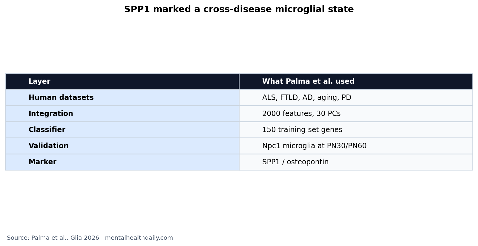
- The analysis used high-dimensional cell data: researchers integrated 2000 variable features and used the first 30 principal components for clustering and UMAP.1
- Classification was deliberately constrained: the random-forest model retained the 150 most variable training-set genes to classify disease condition on held-out cells.1
- SPP1 had biological follow-through: mouse Npc1 microglia at postnatal day 30 and 60 supported conservation of the disease-associated signal.1
- Clinical translation is premature: a 150-gene classifier in single-cell data is not a blood test, imaging test, or treatment rule.1
Microglia are the brain’s resident immune cells. They prune synapses, clear debris, respond to injury, and can shift into reactive states during neurodegeneration. Single-nucleus RNA sequencing measures gene expression in individual cell nuclei, letting researchers compare cell states that bulk tissue averages would blur.
Palma et al. asked whether different neurodegenerative diseases share a microglial state, not whether all diseases are the same. That distinction keeps the result useful: common inflammatory programs can coexist with disease-specific triggers and anatomy.
5 Neurodegenerative Contexts Shared a Microglial Program
The analysis pulled human single-nucleus RNA-sequencing datasets from ALS, frontotemporal dementia, Alzheimer’s disease, aging, and Parkinson’s disease, then selected microglial populations for integrated analysis.1 After preprocessing, the workflow used 2000 variable features, 30 principal components, Harmony integration, and marker-based cluster annotation.
Frontotemporal dementia refers to disorders that damage frontal and temporal brain networks, often changing behavior, language, or executive function. ALS damages motor neurons. Alzheimer’s and Parkinson’s have different hallmark proteins and symptoms. Finding overlap across those conditions therefore points to a shared immune-cell response rather than a shared diagnosis.
Principal components are compressed mathematical summaries of variation in high-dimensional data. Using 30 principal components means the researchers let many gene-expression signals shape clustering without treating every gene as equally informative. UMAP is a visualization method that places cells with similar expression profiles near each other; it helps display structure but does not prove biology by itself.
The important methodological point is that Palma et al. tried to reduce dataset noise before interpreting disease states. Public single-cell datasets differ in tissue source, disease stage, laboratory workflow, sequencing chemistry, and annotation. Harmony integration and microglia-only refinement were attempts to compare like with like.
SPP1 Was the Marker, Not the Whole Mechanism
SPP1 encodes osteopontin, an immune and extracellular-matrix-associated protein. In microglia, SPP1 is often treated as a marker of reactive or disease-associated states rather than a standalone disease cause.
The study highlighted SPP1 because the cross-disease program aligned with inflammatory and neurodegenerative processes, and because Spp1 expression was validated in primary microglia from a Niemann-Pick type C mouse model. Niemann-Pick type C is a lipid-storage neurodegenerative disorder; in mice, it produces microglial activation, lysosomal dysfunction, and progressive neurological decline.
That validation step matters because machine-learning features can sometimes separate labels without mapping neatly onto biology. Here, Spp1 expression localized to disease-relevant regions and appeared in isolated microglia from affected animals, giving the computational signal a biological anchor.1
Osteopontin biology: SPP1 marks more than a visual label on activated microglia. Osteopontin can participate in cell adhesion, migration, immune signaling, and tissue remodeling. In neurodegeneration, those functions can be double-edged: phagocytic cleanup may help remove debris, while chronic inflammatory activation may damage synapses or neurons.
That double-edged biology is why “SPP1-positive” should not be read as automatically good or bad. A microglial state can be protective at one stage and harmful at another, or useful in one disease compartment and destructive in another. The paper identifies a conserved state; it does not assign one universal clinical meaning to that state.
The 150-Gene Classifier Shows State Separation, Not Diagnosis
The random-forest model used the 150 most variable genes from the training set and tested classification on held-out cells. Random forest means an ensemble machine-learning model that combines many decision trees; it can classify patterns but does not automatically identify causal mechanisms.
Important limit: classifying disease labels from purified microglial transcriptomes is not the same as diagnosing a living patient. Brain tissue, single-cell sequencing, dataset harmonization, and curated cell selection are far from a routine clinical assay.
The stronger implication is research-facing: SPP1-positive microglial states may be a useful cross-disease readout for testing whether interventions calm or redirect neurodegeneration-associated inflammation.
What classification adds: if a 150-gene model can separate disease conditions from controls in held-out cells, then microglial disease states contain enough information to be recognized computationally. That does not require each disease to have a perfectly unique microglial pattern. Shared and disease-weighted components together can carry signal.
Feature importance also helps generate mechanistic candidates. Genes that repeatedly help classification can be followed up in animal models, organoids, or human postmortem validation. SPP1 is attractive in that sense because prior Alzheimer’s model work already connected it to microglial phagocytosis and synaptic engulfment.
Prior Disease-Associated Microglia Work Predicted This Direction
Deczkowska et al. framed disease-associated microglia as a recurring immune response across neurodegeneration.2 Palma et al. extends that idea with integrated human transcriptomics and explicit cross-condition classification.
De Schepper et al. linked SPP1 to microglial phagocytic states and synaptic engulfment in Alzheimer’s model systems.3 That adjacent work makes the SPP1 finding less like a random marker and more like part of a known reactive-microglia axis.
Bright et al. reviewed neuroinflammation in frontotemporal dementia, where microglia and other immune pathways can shape disease context beyond classic protein-aggregation narratives.4 Together, the literature supports a calibrated thesis: neurodegenerative diseases differ, but their immune-cell responses can converge.
Evidence Strength and Limits
Supported: integrated human single-nucleus datasets reveal a conserved microglial transcriptional program across multiple neurodegenerative contexts, and SPP1 is a strong marker within that program.
Not supported: using SPP1 alone as a clinical biomarker, treating SPP1 as universally harmful, or assuming microglia behave identically in Alzheimer’s disease, Parkinson’s disease, ALS, FTLD, and aging. The study’s mouse validation strengthens biological plausibility, but it remains preclinical and cell-state focused.
Best use of the result: future neurodegeneration studies can ask whether a therapy changes the SPP1-positive state, whether that change tracks neuronal survival or synaptic preservation, and whether the same state appears in cerebrospinal fluid or imaging-adjacent biomarkers. Those are testable next steps. They are stronger than a vague claim that inflammation matters in all brain diseases.
For readers, the practical takeaway is that microglia are not background support cells. They carry disease-state information across several neurodegenerative disorders, and SPP1 is one of the clearer handles on that shared immune-cell response.
Therapeutic caution: a shared microglial state does not automatically mean one anti-inflammatory drug would work across ALS, FTLD, Alzheimer’s disease, Parkinson’s disease, and aging. Timing, region, disease stage, and the surrounding protein pathology still matter. A therapy that suppresses debris clearance could backfire if the SPP1-positive state is partly compensatory in that context.
The better near-term use is biomarker-guided experimentation. Researchers can compare whether models with different initiating pathologies converge on the same SPP1-positive program, then test whether changing that program improves neuronal outcomes or merely changes a marker. That separates a useful therapeutic target from a useful disease-state label.
Human validation also needs spatial context. A microglial transcriptomic state near plaques, degenerating motor pathways, or vulnerable frontal networks may have different meaning than the same marker in less affected tissue. Combining single-cell data with spatial transcriptomics and neuropathology would make the SPP1 signal more interpretable.
That spatial step would also help distinguish local injury response from a brain-wide inflammatory state.
It would also make negative findings easier to interpret. If a future study does not find SPP1 enrichment in blood or cerebrospinal fluid, that may mean the marker is too tissue-localized for peripheral testing rather than biologically irrelevant.
Assay choice is not a detail. Single-nucleus RNA sequencing can detect cell-state differences that bulk tissue, serum cytokines, or routine inflammatory markers may miss. A weak peripheral SPP1 result would not necessarily contradict a strong microglial signal inside degenerating tissue; it would show that the measurement layer changed.
That is why the article’s safest interpretation is translational, not diagnostic: SPP1 helps name a conserved disease-state program that future assays may or may not capture outside brain tissue.
Questions About SPP1 Microglia
Is SPP1 a blood biomarker for dementia or Parkinson’s disease?
Not from this paper. The signal came from microglial transcriptomics and mouse microglia validation. A blood biomarker would require separate assay and clinical validation.
Does shared microglial activation mean the diseases have one cause?
No. Shared immune-cell states can appear downstream of different disease triggers, including amyloid, tau, alpha-synuclein, motor-neuron injury, lysosomal stress, or aging-related tissue damage.
How can researchers use the SPP1 signal now?
The most immediate use is research stratification: SPP1-positive microglial states can help compare disease models and test whether experimental treatments alter a conserved neuroinflammatory program.
References
- Palma A, Stefanelli R, Trenta F, et al. A cross-disease microglial transcriptional program characterizes neurodegeneration and highlights SPP1 as a biomarker. Glia. 2026. doi:10.1002/glia.70163
- Deczkowska A, Keren-Shaul H, Weiner A, Colonna M, Schwartz M, Amit I. Disease-associated microglia: a universal immune sensor of neurodegeneration. Cell. 2018. doi:10.1016/j.cell.2018.05.003
- De Schepper S, Ge JZ, Crowley G, et al. Perivascular cells induce microglial phagocytic states and synaptic engulfment via SPP1 in mouse models of Alzheimer’s disease. Nature Neuroscience. 2023. doi:10.1038/s41593-023-01257-z
- Bright F, Werry EL, Dobson-Stone C, et al. Neuroinflammation in frontotemporal dementia. Nature Reviews Neurology. 2019. doi:10.1038/s41582-019-0231-z