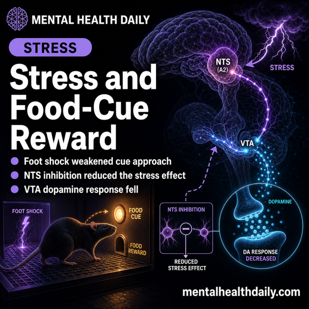
A 2026 rat study found that foot-shock stress transiently reduced food-cue approach during the first 2 conditioned-stimulus trials (t = 2.807; P < 0.05), and inhibiting the nucleus of the solitary tract weakened that stress effect.1 The useful finding is selective: stress used an NTS-linked route to dampen cue-evoked VTA dopamine, while outcome-specific satiety reduced cue behavior through a different route.
Research Highlights
- Stress changed early cue responding: foot shock produced a stress x trial interaction (F(7,70) = 2.867; P < 0.05) and reduced CS+ elevation during the first 2 trials.
- Satiety had a later cue effect: sucrose prefeeding reduced cue response in the last 2 CS+ trials (F(2,18) = 6.141; P < 0.01), separating satiety timing from acute stress timing.
- NTS inhibition weakened stress suppression: rats receiving CNO plus foot shock showed higher early CS+ elevation than vehicle plus foot shock (F(2,14) = 7.587; P < 0.05).
- A2 neuron activation was sufficient: chemogenetic activation of NTS A2 neurons reduced CS+ elevation in hM3Dq rats (F(1,4) = 19.516; P < 0.05).
- VTA dopamine was the downstream signal: NTS A2 activation reduced CS+ VTA dopamine AUC (t = 3.270; P = 0.01), while lateral hypothalamus activity did not shift.
Nucleus of the solitary tract (NTS) is a hindbrain hub that receives visceral and interoceptive information from the body. In feeding behavior, it sits where stress physiology, gut-derived signals, and reward-cue processing can plausibly meet.
A2 neurons are noradrenergic NTS neurons, meaning they use norepinephrine signaling and respond strongly to stress-related input. Yap et al. tested whether activating or inhibiting this population could change conditioned approach to a sucrose-predictive cue.
Foot-Shock Stress Suppressed the First 2 Food-Cue Trials
The behavioral setup was a Pavlovian appetitive conditioning task. Rats learned that one 15-second conditioned stimulus predicted sucrose and another 15-second stimulus did not. The response measure was elevation in food-magazine entries during the cue, compared with the 15 seconds before the cue.
After 30 minutes of 0.6 mA foot shock, stress did not produce one uniform drop across the entire test. Instead, the stress effect was concentrated early. The stress x trial interaction reached significance (F(7,70) = 2.867; P < 0.05), and the first 2 CS+ trials were lower after foot shock (t = 2.807; P < 0.05). The last 2 CS+ trials were not lower (t = −0.534; P = 0.605).
Timing separates stress from satiety: sucrose prefeeding also reduced cue-induced appetitive behavior, but its clearer effect appeared later in the session. That pattern argues against a single “less hungry, less cue response” explanation.
Outcome-Specific Satiety Did Not Use the Same NTS Route
Outcome-specific satiety means the animal was selectively fed the same reward that the cue predicted, reducing the value of that exact outcome. Here, sucrose prefeeding lowered CS+ responding compared with ad libitum feeding, while chow refeeding did not produce the same effect.
Satiety in this task was reward-specific fullness: eating sucrose reduced the motivational pull of a sucrose cue. Sucrose prefeeding lowered late CS+ elevation, while latency to enter the magazine did not significantly change (F(2,18) = 3.966; P = 0.08).
NTS inhibition did not reverse the sucrose-prefeed suppression. Stress and satiety both pushed cue behavior down, but the NTS requirement appeared stress-selective in this experiment.
NTS Inhibition Attenuated Stress-Induced Cue Suppression
Yap et al. used inhibitory DREADD chemogenetics to silence NTS neurons before foot shock. DREADDs are engineered receptors that let researchers turn selected neurons up or down with a drug-like ligand; in this case, CNO was used before stress exposure.
The strongest NTS result came from the first 2 conditioned cues after foot shock. During those early trials, condition affected CS+ elevation (F(2,14) = 7.587; P < 0.05). Vehicle plus foot shock reduced CS+ elevation compared with no foot shock, but CNO plus foot shock attenuated that suppression.
- No-stress control: cue approach remained intact.
- Vehicle plus foot shock: stress lowered early CS+ elevation.
- CNO plus foot shock: NTS inhibition weakened the stress-induced drop.
A control experiment in animals without hM4Di expression kept the interpretation honest. CNO failed to rescue cue responding in those animals, supporting a receptor-dependent NTS inhibition effect rather than a nonspecific CNO artifact.
A2 Neuron Activation Reduced Cue-Evoked VTA Dopamine
Activating NTS A2 neurons without foot shock mimicked part of the stress effect. In hM3Dq rats, CNO reduced CS+ elevation compared with vehicle (F(1,4) = 19.516; P < 0.05), while CS- responding did not show the same target-specific drop.
Ventral tegmental area (VTA) dopamine neurons help assign motivational value to cues that predict rewards. Fiber photometry showed that NTS A2 activation reduced CS+ VTA dopamine activity (t = 3.270; P = 0.01), without reducing activity during CS-, pellet delivery, or pellet retrieval.
Foot shock produced a similar dopamine pattern. Stress suppressed VTA dopamine during CS+ (t = 2.739; P < 0.05), supporting the idea that acute stress and NTS A2 activation converge on the cue-evoked dopamine response.
Lateral Hypothalamus Activity Did Not Explain the Main Effect
Lateral hypothalamus is a feeding and arousal region that can influence cue-induced food seeking. Because NTS A2 neurons project to multiple feeding-related areas, the study also measured lateral hypothalamus activity during the same conditioning task.
Activation of NTS A2 neurons reduced cue approach, but it did not significantly change lateral hypothalamus activity during CS+ (t = −1.228; P = 0.251) or CS- (t = 0.664; P = 0.523). The experiment therefore points more strongly toward VTA dopamine than toward a broad lateral-hypothalamus suppression signal.
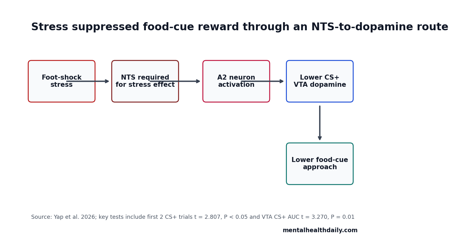
Circuit implication: acute stress may suppress food-cue reward by recruiting hindbrain stress-responsive neurons that lower dopamine response to the cue itself. That is narrower than saying stress globally suppresses feeding circuits.
Rodent Circuit Evidence Is Mechanistic, Not a Human Treatment Claim
Evidence-strength note: this was an animal-only circuit study using foot shock, chemogenetics, and fiber photometry. It supports mechanism direction inside a controlled rat task. It does not establish a treatment for human stress eating, binge eating, obesity, or addiction.
Adjacent work makes the result plausible. Rodent studies have shown that chronic social defeat and acute foot shock can suppress conditioned food-cue responses. Other NTS-focused work has implicated GLP-1, oxytocin, and noradrenergic signaling in reducing conditioned food seeking or food-cue motivation.2
VTA dopamine work also fits the direction. Food-predictive cues normally evoke dopamine signals that help drive approach. A stress-responsive hindbrain pathway that lowers that cue-evoked dopamine signal gives the behavioral effect a concrete circuit route.3
The Circuit Separates Cue Value From General Movement
The behavioral measure was not simple locomotion or total food intake. It was cue-triggered magazine entry during a 15-second conditioned stimulus, compared with the 15 seconds before that cue. That design targets the learned motivational pull of the cue rather than asking whether stressed rats were globally inactive.
Early-trial specificity matters: foot shock reduced CS+ elevation in the first 2 trials, then the effect faded by the last 2 trials. A broad motor-suppression account would predict a more uniform session-wide deficit. The time course fits a transient stress-state effect on cue value better than a simple inability to approach the food cup.
NTS inhibition sharpened the inference: if NTS activity were irrelevant, inhibiting it should not have protected cue response after foot shock. Instead, CNO plus foot shock left higher early CS+ elevation than vehicle plus foot shock, placing NTS activity upstream of the stress-induced behavioral change.1
Dopamine readout closed the loop: NTS A2 activation reduced cue-evoked VTA dopamine AUC, the signal expected to carry learned food-cue value. The lateral hypothalamus result helped narrow the pathway further because feeding-related nodes did not all move with the manipulation, making the dopamine effect more pathway-specific.
The human translation problem is that stress can increase eating in some settings and suppress it in others. This rat task isolates one acute, aversive stressor and one learned sucrose cue. It does not model chronic stress, dieting history, binge-eating risk, trauma exposure, or food availability. The useful inference is therefore circuit-level: an acute stress state can reduce the reward value of a food-predictive cue through an NTS-to-dopamine route.
Future work should test whether the same pathway changes when the stressor is social, chronic, or controllable; whether females show the same time course; and whether high-calorie foods behave differently from sucrose pellets. Those experiments would decide whether the NTS A2 signal is a general stress-appetite pathway or a narrower acute-threat mechanism.
The timing result also argues for testing recovery dynamics. If cue response normalizes within the same session, the pathway may be most relevant to short-lived stress states. If repeated stress makes the dopamine suppression durable, the same NTS route could become relevant to longer-term changes in motivation and feeding behavior.
Satiety is the internal comparator: the experiment is stronger because it tested 2 ways to suppress cue-driven approach. Sucrose prefeeding changed behavior later in the session and did not depend on the same NTS manipulation. That contrast keeps the stress result from becoming a generic “less motivation” story.
For addiction and eating-disorder interpretation, the safer takeaway is about state control of cue response. The same learned cue can have different motivational weight depending on internal state. Acute stress, satiety, and dopamine signaling can shift that weight, but they do not all have to use the same circuit route or produce the same time course.
Questions About Stress, Food Cues, and NTS Dopamine Circuits
Did stress make the rats stop responding to all cues?
No. The clearest stress effect appeared during the first 2 CS+ trials after foot shock, and the last 2 CS+ trials were not significantly reduced.
Did satiety and stress use the same mechanism?
Not in this experiment. NTS inhibition attenuated the stress effect, while sucrose-prefeed suppression remained NTS-independent under the tested conditions.
Does this prove a stress-eating treatment target?
No. The result is a rat circuit finding. It identifies a plausible stress-to-dopamine pathway that future work can test in broader feeding and reward models.
References
- Yap JA, Gu YH, Vuong C, Power JM, Ong ZY. Nucleus of the solitary tract regulation of cue-induced appetitive behaviors via midbrain dopamine neurons. Neuropsychopharmacology. 2026. https://doi.org/10.1038/s41386-026-02417-y
- PubMed search: nucleus solitary tract GLP-1 oxytocin cue induced food seeking. https://pubmed.ncbi.nlm.nih.gov/?term=nucleus+solitary+tract+GLP-1+oxytocin+cue+induced+food+seeking
- PubMed search: VTA dopamine food predictive cue conditioned approach. https://pubmed.ncbi.nlm.nih.gov/?term=VTA+dopamine+food+predictive+cue+conditioned+approach
- PubMed search: stress foot shock cue induced feeding behavior rats. https://pubmed.ncbi.nlm.nih.gov/?term=stress+foot+shock+cue+induced+feeding+behavior+rats