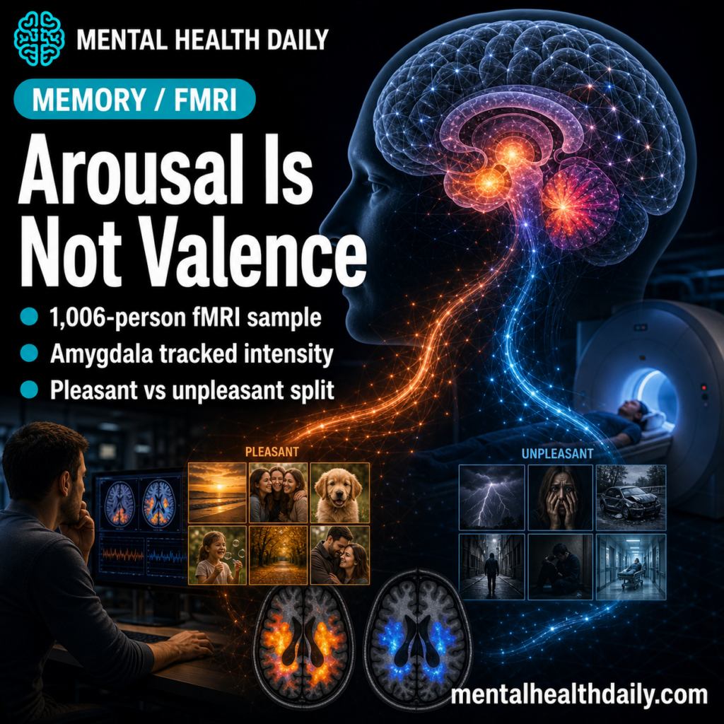
A 2026 fMRI study of 1,006 healthy young adults found that emotional pictures were remembered better than neutral pictures, but the brain signal split after arousal was modeled: amygdala and insula effects dropped out, while negative and positive valence kept separate cortical encoding patterns.1
Research Highlights
- Emotional pictures had the recall advantage: in the full behavioral sample of 1,591 adults, participants recalled 12.2 positive pictures and 11.4 negative pictures on average, compared with 7.2 neutral pictures out of 72.1
- Positive pictures beat negative pictures despite lower arousal: positive recall exceeded negative recall by 0.8 pictures, even though negative pictures had higher subjective arousal ratings, 2.36 vs. 1.94 on a 3-point arousal scale.1
- Amygdala and insula looked arousal-sensitive: emotional > neutral encoding initially included bilateral insula and left amygdala, but those clusters were no longer significant after subjective arousal was modeled.1
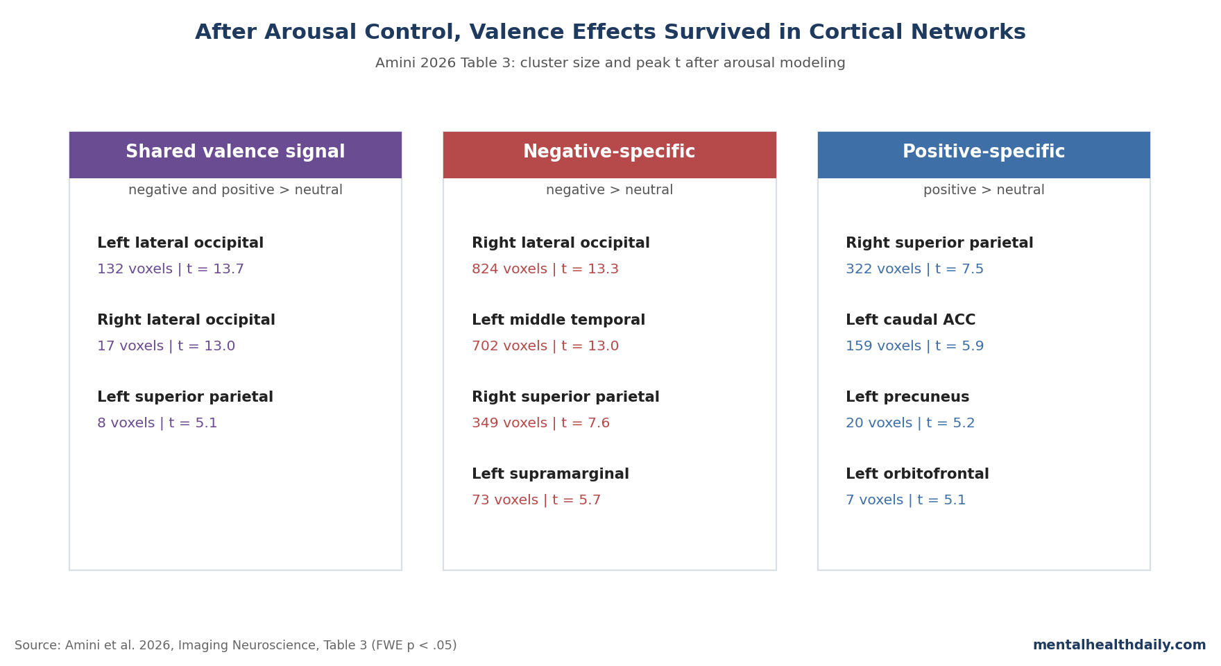
- Negative valence survived in visual-sensory regions: after arousal control, negative-specific memory encoding included a right lateral occipital cluster of 824 voxels and a left middle temporal cluster of 702 voxels.1
- Positive valence survived in midline-control regions: positive-specific encoding included a right superior parietal cluster of 322 voxels, left caudal anterior cingulate cluster of 159 voxels, and precuneus/orbitofrontal clusters smaller than the 322-voxel superior parietal cluster.1
Valence means whether an emotional event is pleasant or unpleasant. Arousal means how activating or intense the event feels.
They often travel together in ordinary life, especially when an unpleasant event is also intense, but they are not the same variable.
Amini et al. used a large picture-encoding fMRI dataset to separate those variables more cleanly than most older emotional-memory studies could. Participants viewed negative, neutral, and positive pictures during scanning, rated each picture for valence and arousal, then completed an unannounced free-recall test around 10 minutes later.1
Positive Pictures Were Remembered Most, Even With Lower Arousal
The behavioral result is easy to miss because the brain analysis is the flashier part. Across 1,591 participants, total free recall averaged 30.8 of 72 pictures.
Positive pictures were remembered most often at 12.2 pictures on average, followed by negative pictures at 11.4 and neutral pictures at 7.2.1
Both emotional categories strongly beat neutral recall. Negative vs. neutral recall produced Cohen’s d = 1.26, and positive vs. neutral recall produced Cohen’s d = 1.49, both with p < .001.
The positive-over-negative difference was smaller, d = -0.25, but still statistically reliable.1
That ordering matters because negative pictures were more arousing than positive pictures: 2.36 vs. 1.94 on the study’s 1-to-3 subjective arousal scale. If arousal alone explained emotional memory, negative pictures should have had the clearer recall advantage.
Instead, the study found a small positive-memory edge, suggesting that memorability, nameability, semantic richness, retrieval accessibility, and valence-specific encoding processes can push memory beyond raw activation level.1
Amygdala and Insula Dropped Out After Arousal Control
Subsequent-memory analysis asks which brain activity during encoding predicts later remembering. In the first whole-brain model, Amini et al. compared emotional difference-in-memory effects with neutral difference-in-memory effects.
Emotional > neutral successful encoding produced large occipito-temporal clusters, anterior cingulate cortex, insula, and left amygdala activation, using family-wise-error correction at p < .05.1
That first pass mostly agrees with older fMRI work: emotional memory involves salience, visual processing, and emotion-sensitive regions rather than only a single “memory center.” McGaugh’s consolidation model made the amygdala central to emotionally arousing memories, and Kensinger and Corkin’s 2004 study proposed 2 routes: a high-arousal amygdala-hippocampal route and a more elaborative prefrontal route for lower-arousal emotional material.2,9
The more important move came next. Amini et al. used parametric modulation, a model that entered each participant’s subjective arousal rating before the memory term, so the remaining memory signal represented encoding variance not explained by arousal.
After that control, amygdala and insula clusters were no longer significant. The paper’s own discussion states the interpretation directly: these regions appeared to track emotional arousal rather than valence-specific memory enhancement.1
Negative Valence Survived as Visual-Sensory Encoding
After arousal control, negative emotional memory enhancement kept its strongest signal in posterior visual and sensory-attentional territory. Table 3 reported a right lateral occipital cluster of 824 voxels with peak t = 13.3, a left middle temporal cluster of 702 voxels with peak t = 13.0, a right superior parietal cluster of 349 voxels with peak t = 7.6, and a left supramarginal cluster of 73 voxels with peak t = 5.7.1
That pattern fits a long-running idea in emotional-memory research: negative material often gets encoded with sharper sensory detail. Mickley Steinmetz and Kensinger found that valence and arousal shaped neural activity leading to later memory, while later connectivity work showed that arousal effects on the emotional-memory network depended on whether stimuli were negative or positive.4,5
Amini et al. sharpened the claim by showing that negative-valence clusters survived even after subjective arousal was statistically accounted for. That does not prove the regions cause negative memory enhancement.
It does show that the surviving negative-memory signal was more than the amygdala/insula arousal signature wearing a valence label.
Positive Valence Survived in ACC, Frontal, and Parietal Networks
Positive emotional memory enhancement looked different after arousal control. The largest positive-specific cluster was right superior parietal cortex, 322 voxels with peak t = 7.5.
Smaller positive-specific clusters included left caudal anterior cingulate cortex, 159 voxels with peak t = 5.9; left precuneus, 20 voxels with peak t = 5.2; and left lateral orbitofrontal cortex, 7 voxels with peak t = 5.1.1
Anterior cingulate cortex is a midline brain region involved in attention, emotion regulation, action selection, and reward-related processing. Orbitofrontal cortex helps represent value, preference, and reward.
Their appearance in positive-memory encoding fits earlier work suggesting that positive memory relies more on elaborative, prefrontal, and value-linked processes than negative memory does.6,7
The result is not a clean “positive equals reward cortex” slogan. Positive-specific memory enhancement also involved parietal and precuneus regions, and several positive clusters were small.
The better read is anatomical patterning: negative remembered pictures leaned toward sensory-attentional visual systems, while positive remembered pictures leaned toward midline, frontal, parietal, and default-mode-related systems after arousal was modeled.
Free Recall Did Not Reproduce Hippocampal Meta-Analysis Results
One of the strongest calibration points is what the study did not find. Dahlgren et al.’s 2020 coordinate-based meta-analysis linked successful emotional episodic encoding and retrieval to medial temporal regions, including hippocampal and parahippocampal areas, along with visual and salience-related regions.3
Amini et al. replicated part of that landscape but not the medial-temporal piece. Emotional > neutral successful encoding included visual cortex, insula, amygdala, and anterior cingulate effects, yet hippocampal/parahippocampal activations were absent in the main emotional-memory contrast.1
The authors’ workup is plausible: most studies in the meta-analysis used recognition, while Amini et al. used free recall. Recognition can be supported by familiarity and low-confidence memory signals.
Free recall demands self-initiated retrieval and may define “forgotten” more harshly, because a picture that is not freely recalled might still be recognized later. That design difference can weaken medial-temporal remembered-vs-forgotten contrasts even when hippocampal systems remain essential for memory more generally.
Neutral Memory Used a Different Encoding Strategy
Neutral pictures recruited their own encoding pattern. After arousal control, neutral memory showed stronger activation than emotional memory in left fusiform cortex, right parahippocampal cortex, and right precuneus for the shared neutral > negative and neutral > positive contrast.
Additional neutral > negative clusters appeared in frontal, inferior parietal, lingual, and parahippocampal regions.1
This fits a practical memory principle: when a picture does not carry emotional salience, the brain may lean harder on scene processing, object recognition, attention control, and deliberate elaboration. A neutral room, building, or object has to be encoded as a describable scene.
A negative or positive picture may recruit salience and valence systems before the person tries to name it.
The study checked whether remembered content categories were driving the neutral result. A trial-level mixed-effects model found no evidence that content category changed the memory relationship differently across valence categories, with no valence-by-content interaction, F = 0.84 and p > .05.1
Limits of This fMRI Decomposition
The study is unusually large for task fMRI, but it is still a healthy-young-adult encoding study. It does not show how these networks behave in post-traumatic stress disorder, depression, dementia, medication exposure, or autobiographical trauma memory.
Arousal measurement was coarse. Participants rated arousal on a 3-point scale. That supports basic fMRI modeling, but it is not the same as skin conductance, heart-rate variability, pupil response, cortisol, or noradrenergic activity.
Valence was not modeled symmetrically. Arousal could be entered as an ordinal parametric modulator, but valence was categorical. That means the researchers could estimate valence-specific memory effects after controlling for arousal, but they could not run the exact mirror question of arousal effects after controlling for valence in the same way.
Exclusions shaped the fMRI sample. The behavioral sample included 1,591 participants, but the group-level fMRI analysis required at least 5 remembered pictures per valence category and behavioral emotional-memory enhancement for both negative and positive categories. That yielded 1,006 participants.
The arousal-control model used 792 participants because some arousal ratings were missing.1
Brain activation is not intervention evidence. The findings refine models of emotional encoding. They do not prove that targeting anterior cingulate cortex, lateral occipital cortex, or amygdala activity will improve therapy or prevent intrusive memories.
Questions About Valence, Arousal, and Emotional Memory
Did the study prove the amygdala is unimportant for emotional memory?
No. The first emotional > neutral model included amygdala activity, and decades of animal, lesion, pharmacological, and human fMRI work support an amygdala role in emotionally arousing memory.2,9
Amini et al. showed something narrower: in this dataset, amygdala and insula effects no longer survived after subjective arousal was modeled.1
Why did positive pictures have better recall than negative pictures?
Positive-memory interpretation: positive pictures had a small recall advantage, 12.2 vs. 11.4 pictures, even though negative pictures were rated more arousing. The study cannot reduce that difference to one mechanism, but it points away from a pure arousal explanation and toward stimulus memorability, retrieval accessibility, nameability, and valence-specific encoding routes.1
How should “controlling for arousal” be read here?
The model used each participant’s subjective arousal rating as the first parametric modulator, then tested whether memory-related brain activity remained after that arousal-linked variance was accounted for. It is a statistical control, not a physiological clamp on arousal.
Does this directly explain PTSD flashbacks?
Not directly. PTSD involves real autobiographical trauma, long-term consolidation, sleep, stress hormones, threat learning, and repeated retrieval.
A 10-minute free-recall fMRI task in healthy young adults can refine mechanisms, but it should not be sold as a PTSD biomarker.
Why is the missing hippocampal signal important?
It prevents overreading the result as a simple amygdala-hippocampus story. Amini et al. suggest that free recall, picture-only stimuli, and how forgotten trials are defined may explain why their main contrast did not reproduce the hippocampal/parahippocampal signal reported in recognition-heavy meta-analytic work.1,3
What is the most useful clinical takeaway?
Do not treat “emotional memory” as one mechanism. High-arousal salience, negative sensory detail, positive elaboration, neutral scene processing, and later consolidation can all matter.
The Amini study helps separate those layers, but treatment claims still require clinical trials rather than fMRI pattern matching.
References
- Neural correlates of emotional memory enhancement: the role of valence and arousal. Amini E et al. Imaging Neuroscience. 2026;4:1213. doi:10.1162/imag.a.1213
- Two routes to emotional memory: distinct neural processes for valence and arousal. Kensinger E, Corkin S. Proceedings of the National Academy of Sciences. 2004;101(9):3310–3315. doi:10.1073/pnas.0306408101
- Neural correlates of successful emotional episodic encoding and retrieval: an SDM meta-analysis of neuroimaging studies. Dahlgren K et al. Neuropsychologia. 2020;143:107495. doi:10.1016/j.neuropsychologia.2020.107495
- The effects of valence and arousal on the neural activity leading to subsequent memory. Mickley Steinmetz KR, Kensinger EA. Psychophysiology. 2009;46(6):1190–1199. doi:10.1111/j.1469-8986.2009.00868.x
- The effect of arousal on the emotional memory network depends on valence. Mickley Steinmetz KR et al. NeuroImage. 2010;53(1):318–324. doi:10.1016/j.neuroimage.2010.06.015
- Level of processing modulates the neural correlates of emotional memory formation. Ritchey M et al. Journal of Cognitive Neuroscience. 2011;23(4):757–771. doi:10.1162/jocn.2010.21487
- Representing the Good and Bad: fMRI signatures during the encoding of multisensory positive, negative, and neutral events. Thakral PP et al. Cortex. 2022;151:240–258. doi:10.1016/j.cortex.2022.02.014
- Effect of emotional valence on retrieval-related recapitulation of encoding activity in the ventral visual stream. Kark SM, Kensinger EA. Neuropsychologia. 2015;78:221–230. doi:10.1016/j.neuropsychologia.2015.10.014
- The amygdala modulates the consolidation of memories of emotionally arousing experiences. McGaugh JL. Annual Review of Neuroscience. 2004;27:1–28. doi:10.1146/annurev.neuro.27.070203.144157