In an MRI study of 337 young people aged 8 to 18, autistic boys and autistic girls each showed their own cluster of brain structure differences compared with peers of the same sex.1
The boys’ pattern centered on a face-processing region and tracked with social and communication traits; the girls’ pattern centered on a memory hub and did not.
Research Highlights
- Sample: 181 autistic youth (97 boys, 84 girls) and 156 non-autistic youth (78 boys, 78 girls) scanned at 4 US research sites.1
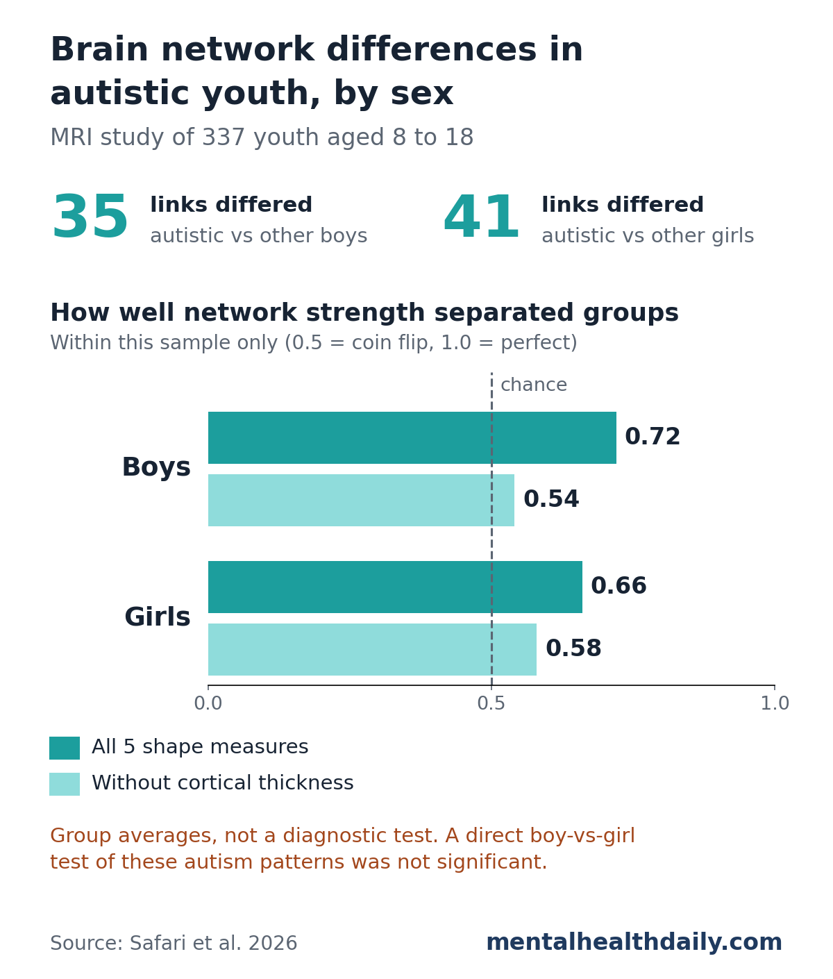
- Boys: 35 brain-region connections differed between autistic and non-autistic boys, mostly as regions being more alike in structure, led by the fusiform gyrus.1
- Girls: 41 connections differed between autistic and non-autistic girls, mostly in the same direction, with the entorhinal cortex as the main hub.1
- No significant boy-girl difference: a direct statistical test of whether autism-related patterns differed by sex came up empty at every threshold tried.1
- Link to autism traits in boys only: the boys’ network correlated with social and communication scores (r = 0.39); the girls’ network showed no such link.1
How Researchers Built Brain “Shape Networks” From MRI Scans
Research by a team led by Nooshin Safari and Javier Rasero at the University of Virginia used standard structural MRI scans, the kind that show the brain’s anatomy rather than its activity. Scans came from the first wave of the NIH Autism Centers of Excellence (ACE) Network, which deliberately recruited equal numbers of autistic boys and girls.1
Most autism research has far more boys than girls, because autism is diagnosed roughly 3 to 4 times more often in males. A meta-analysis by Loomes et al. put the true ratio closer to 3:1 and found signs that girls who meet criteria are less likely to receive a diagnosis.2
Morphology-based connectivity is the key measure here. It does not track nerve fibers or brain activity. Instead, it asks how alike 2 brain regions are in their physical structure.
For each of 68 regions of the cortex (the brain’s wrinkled outer layer), the researchers measured 5 features at thousands of points across the region:1
- Cortical thickness: how thick the gray-matter layer is
- Surface area: how much cortex the region covers
- Sulcal depth: how deep the folds and grooves run
- Mean curvature: how sharply the surface bends
- Gray matter volume: how much gray matter the region holds
A method called MIND (Morphometric Inverse Divergence) then compared the full spread of those measurements between every pair of regions. The more similar 2 regions’ structural “fingerprints,” the stronger their connection. A 2023 validation study across more than 11,000 scans found MIND networks more reliable than an older similarity method and better matched to known nerve-fiber wiring.3
Each participant ended up with a map of 2,278 region-to-region connections. A technique called network-based statistics then searched for clusters of linked connections that differed between groups, after adjusting for age, head size, and scan site.1
Autistic Boys: More Similar Structure Around the Fusiform Gyrus
Compared with 78 non-autistic boys, the 97 autistic boys had one cluster of 35 connections that differed. Nearly all showed regions being more alike in structure in the autistic group. Only 1 weak connection, between 2 temporal-lobe regions, went the other way.1
Key regions in the boys’ cluster:
- Fusiform gyrus (left side): a region on the underside of the brain central to recognizing faces, long studied in autism. It stayed significant even under the strictest statistical cutoff.
- Medial orbitofrontal cortex (left): part of the frontal lobe involved in weighing rewards and social cues.
- Entorhinal cortex and lingual gyrus (right): a memory gateway to the hippocampus and a visual-processing region.
Link to autism traits: within the autistic boys, this network correlated with social and communication scores from the ADI-R, a structured interview with parents about autism traits (r = 0.39, significant after correction). The researchers describe it as a negative correlation: stronger connectivity went with less severe social-communication scores.1
Autistic Girls: A Memory Hub in the Entorhinal Cortex
Compared with 78 non-autistic girls, the 84 autistic girls had a cluster of 41 connections that differed. Again, most showed regions being more alike in structure.1
- Main hub: the entorhinal cortex on both sides of the brain, a gateway for memory and spatial navigation
- Extending to: the inferior parietal lobule, lateral occipital cortex, and the strips of cortex that handle movement and touch, areas involved in combining visual, spatial, and body-sense information
- Fewer decreased connections: mostly involving a groove in the temporal lobe called the superior temporal sulcus, including a link with the fusiform gyrus
Unlike the boys’ network, the girls’ network showed no significant link to autism trait scores. Neither sex’s network linked to scores for anxiety, mood, attention, or behavior problems on a parent checklist.1
Did Autism-Related Brain Patterns Differ Between Boys and Girls?
Not by the study’s formal test. When the researchers tested a sex-by-diagnosis interaction—whether the autism-related differences in boys were statistically different from those in girls—no significant cluster appeared at any of the 3 cutoffs they tried.1
So the boys’ and girls’ clusters look different, centered on different regions, but the data cannot show that the difference between them is real rather than noise. The researchers suggest any true sex difference may be too small to detect with this sample size.1
Not just ordinary sex differences: the team also mapped how non-autistic boys and girls differed from each other, finding a 48-connection network. It shared no connections with the boys’ autism cluster and just 1 with the girls’ cluster.1
Earlier work using a simpler, single-feature method did report sex-dependent network differences in 122 autistic and 122 non-autistic children aged 2 to 4, findings that team called preliminary.4 Age, method, and sample size all differ between the 2 studies.
Why These Brain Differences Are Not a Diagnostic Test
These are group-level averages, and the 2 groups overlapped heavily. Nothing here lets a single scan show whether a child is autistic.
Modest separation, in-sample only: the researchers used each network’s overall strength to see how well it separated autistic from non-autistic youth. The AUC (where 0.5 is a coin flip and 1.0 is perfect) was 0.72 in boys and 0.66 in girls. Those figures came from the same people used to find the networks, which tends to inflate them, and were never checked in a separate group.1
Cortical thickness carried most of the signal: leaving it out dropped the AUC to 0.54 in boys and 0.58 in girls. Removing surface area or volume made almost no difference.1 The researchers themselves state that their approach does not measure diagnostic usefulness and call for machine-learning studies with outside testing.
Autism is diagnosed through developmental history and observation of behavior. Nothing in this study changes that.
Limitations of This Autism MRI Study
- One snapshot in time: the data are cross-sectional, so they cannot show how these patterns develop. Later ACE waves with follow-up scans may answer that.
- Ages 8 to 18 only: brain structure keeps changing into adulthood.
- No formal sex difference: separate patterns in boys and girls are descriptive, since the direct comparison was not significant.
- Modest brain-behavior link: the boys’ correlation was r = 0.39, and girls showed none.
- One type of scan: structural MRI only, without brain activity or white-matter wiring data.
- Research sample: most participants were White and recruited at 4 academic centers.
On the strength side, the sample was balanced by both sex and diagnosis, which most autism imaging samples are not, and both clusters held up after adjusting for scan quality and scan site.1 The fusiform finding in boys also fits a long line of research on face processing in autism.
References
- Safari N, Levy I, Zhang A, et al. Brain morphology network profiles in male and female youth with autism. Cerebral Cortex. 2026;36(9):bhag156. doi:10.1093/cercor/bhag156
- Loomes R, Hull L, Mandy WPL. What is the male-to-female ratio in autism spectrum disorder? A systematic review and meta-analysis. Journal of the American Academy of Child & Adolescent Psychiatry. 2017;56(6):466–474. doi:10.1016/j.jaac.2017.03.013
- Sebenius I, Seidlitz J, Warrier V, et al. Robust estimation of cortical similarity networks from brain MRI. Nature Neuroscience. 2023;26:1461–1471. doi:10.1038/s41593-023-01376-7
- Zielinski BA, et al. Sex-dependent structure of socioemotional salience, executive control, and default mode networks in preschool-aged children with autism. NeuroImage. 2022;257:119252. doi:10.1016/j.neuroimage.2022.119252