Psychopathy, Empathy, and Cortical Structure in 804 Incarcerated Men
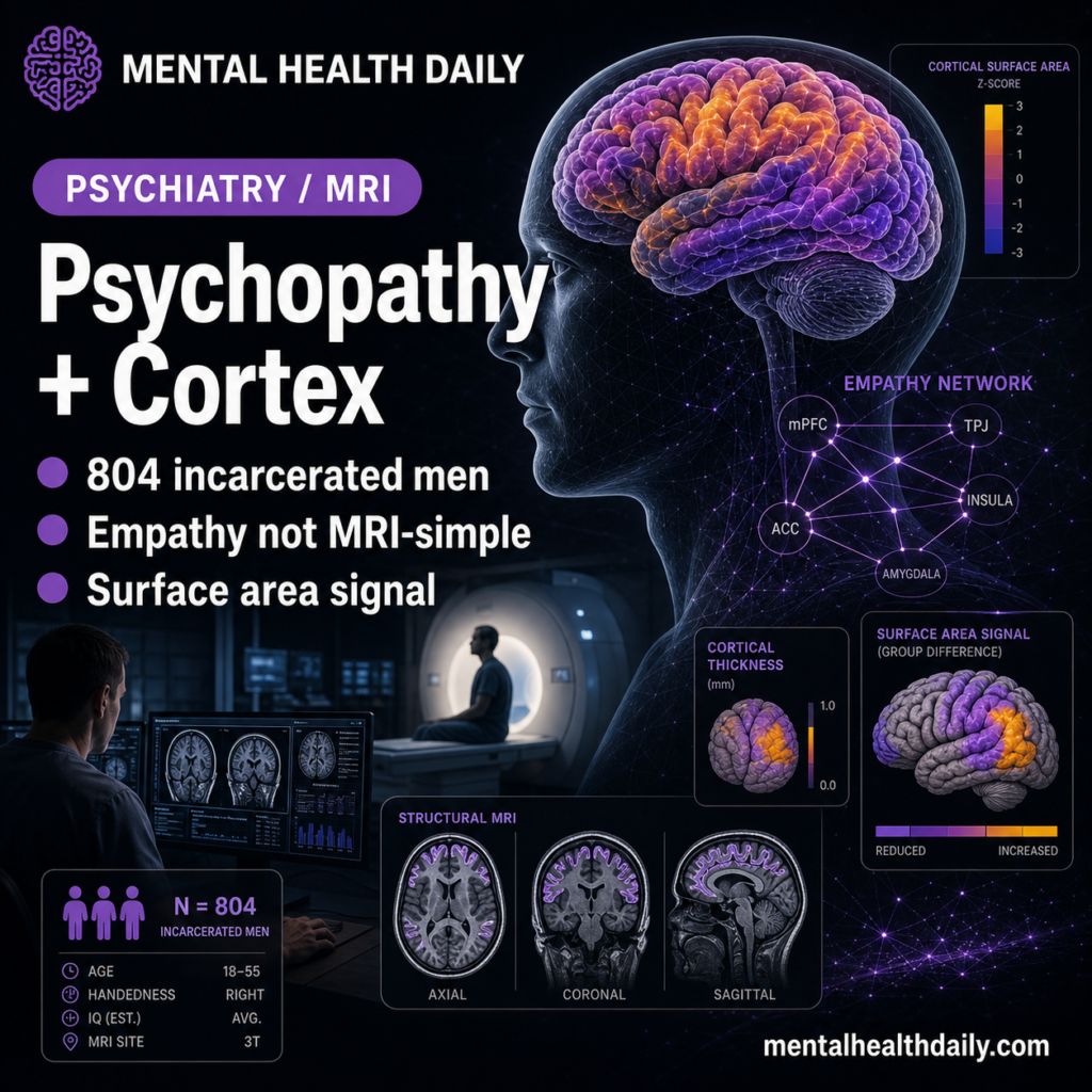
Psychopathy is not a simple “low empathy equals abnormal brain scan” story. In 804 incarcerated men, psychopathy factors tracked empathic concern, perspective taking, cortical thickness, surface area, and cortical organization—but self-reported empathy scores did not map cleanly onto cortical structure.1 Research Highlights The sample was unusually large for forensic MRI. Radecki et al. analyzed 804 …