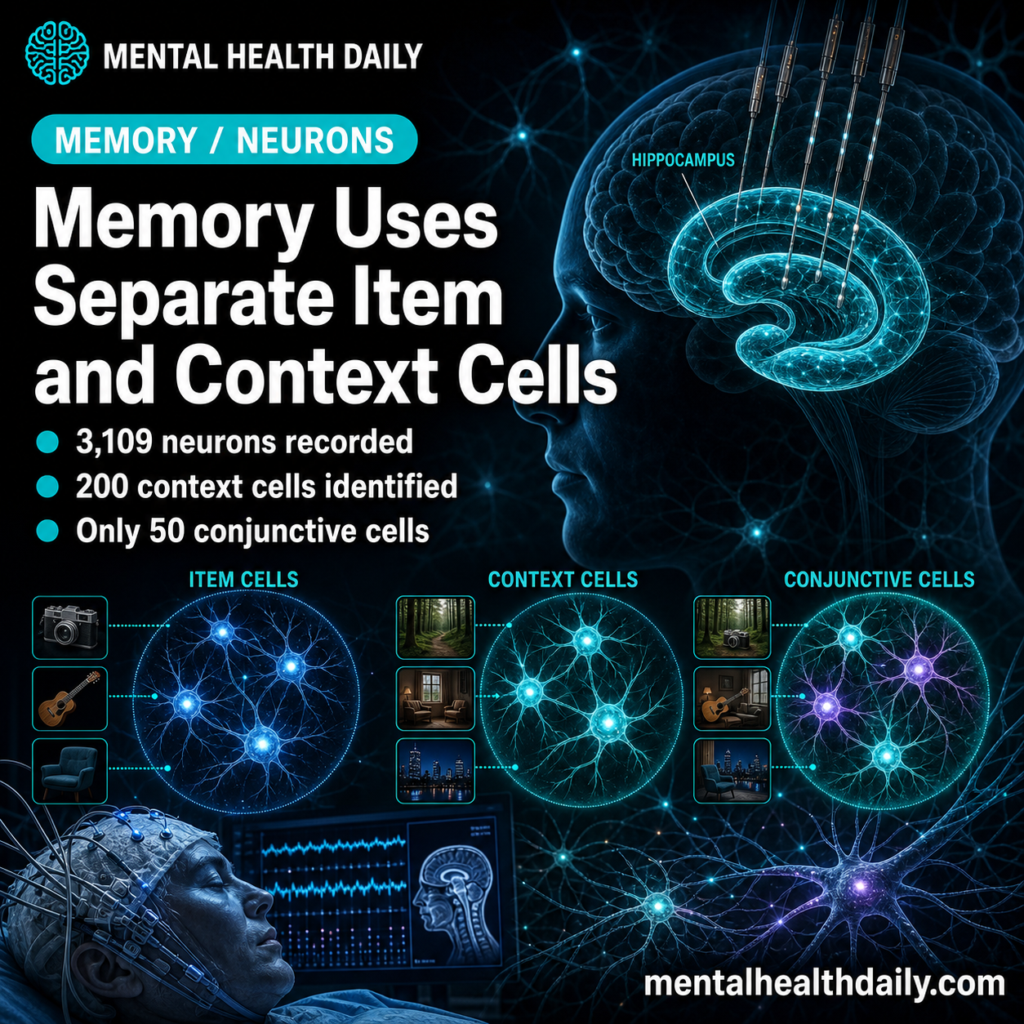
A 2026 Nature single-neuron study recorded 3,109 medial temporal lobe neurons from 16 neurosurgical epilepsy patients and found that human item-context memory mostly used separate item and context populations: 597 stimulus-modulated neurons, 200 context-modulated neurons, and only 50 neurons that encoded specific picture-question combinations.1 The result argues against a simple “one cell stores the whole episode” model of human memory.
Research Highlights
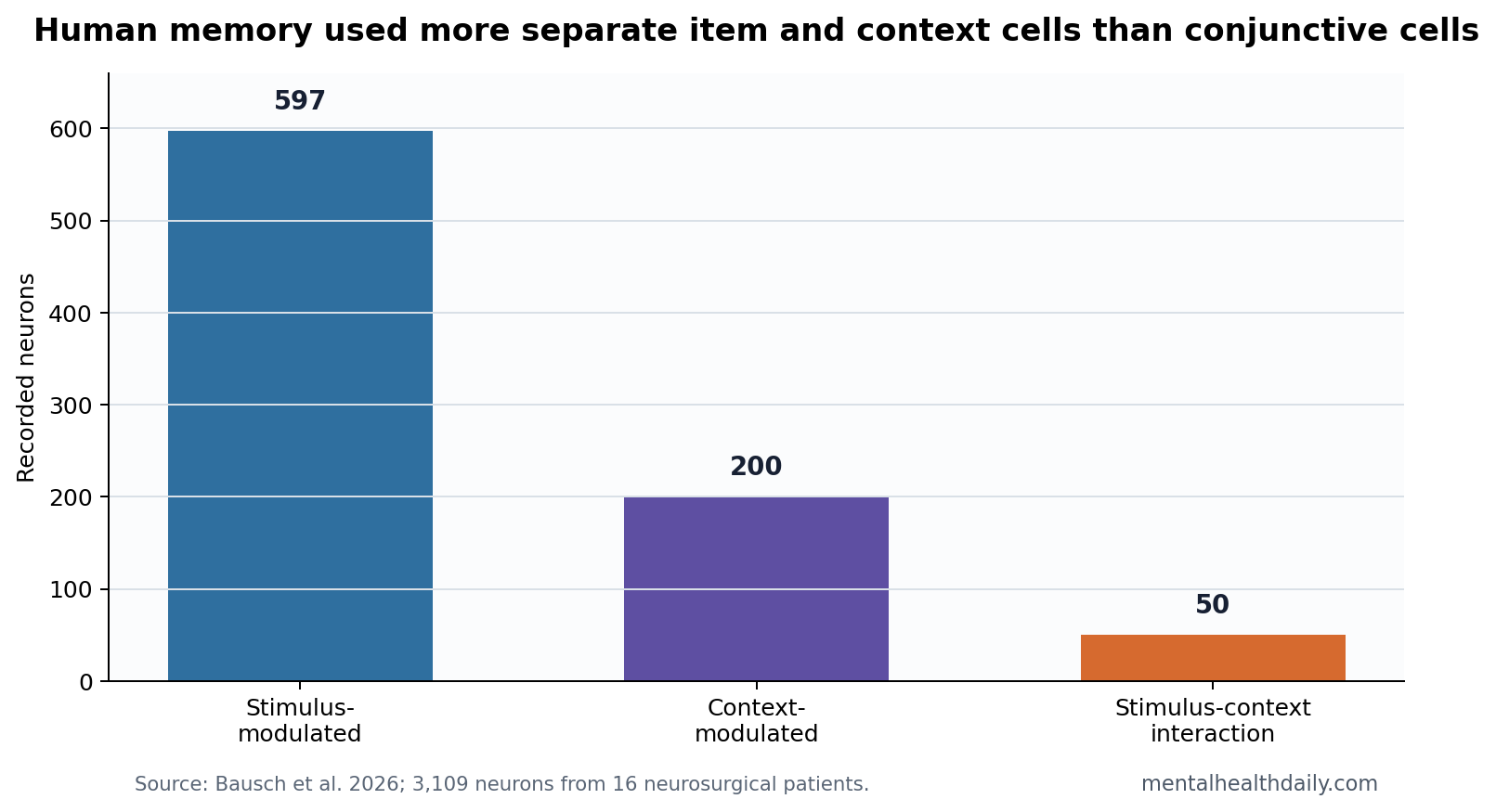
- Separate codes dominated: Bausch et al. identified 597 stimulus-modulated neurons and 200 context-modulated neurons, but only 50 stimulus-context interaction neurons.1
- Context cells were not rare noise: Question-modulated neurons appeared in 2.95% of amygdala neurons, 7.68% of parahippocampal cortex neurons, 5.68% of entorhinal cortex neurons, and 9.42% of hippocampal neurons.1
- Task behavior was valid: Participants’ choices correlated strongly with ground-truth rankings for objective questions, with mean rho = 0.760, p = 6.47 x 10−10.1
- Co-firing carried the binding signal: Stimulus and context neurons were largely separate but covaried with behavioral performance and showed coordinated activity during context reinstatement.1
- Generalization beat rigid binding: The small 50-cell conjunctive population fits a system built around flexible reuse of item and context information, with pattern-separated episode storage as a secondary mechanism.1
Medial temporal lobe refers to a memory-critical brain system that includes the hippocampus, entorhinal cortex, parahippocampal cortex, and amygdala. These areas support declarative memory: facts and events that can be consciously remembered.
Item-context memory means remembering what something was and the setting or rule that made it relevant. In this experiment, the “context” was not a place or life event; it was the comparison question that told participants how to judge 2 pictures, such as which picture was bigger, older, brighter, more expensive, or liked better.
3,109 Human Neurons Split Into Item Cells and Context Cells
Bausch et al. recorded neurons during 49 experimental sessions from patients implanted with depth electrodes for epilepsy monitoring. Participants saw pairs of pictures after a question cue and had to answer according to that cue. A picture could be relevant under several questions, so the task separated picture identity from comparison context.
The central result was anatomical and computationally clean. Many neurons responded to pictures. A smaller but still robust population responded to the question context. Far fewer neurons responded only to one exact picture-question pairing.
- Stimulus-modulated neurons: 597 of 3,109 recorded neurons.
- Context-modulated neurons: 200 of 3,109 recorded neurons.
- Both stimulus and question effects: 73 of 597 stimulus neurons, or 12.23%.
- Conjunctive picture-question cells: 50 of 3,109 neurons, or 1.61%.
Conjunctive coding means a neuron responds to a specific combination, such as one picture under one question, rather than to the picture across questions or to the question across pictures. Rodent hippocampal work often makes this kind of pattern separation central. The human data were more mixed: conjunctive cells existed, but separate reusable codes were more prominent.
Behavioral Performance Kept the Neural Result Interpretable
The task could have failed if participants treated the questions casually or guessed inconsistently. The researchers checked that problem directly. For objective comparisons such as Bigger? and Older? or More expensive?, participant choices correlated strongly with ground-truth rankings, mean rho = 0.760, p = 6.47 x 10⁻¹⁰. Performance also exceeded chance in all but one excluded session and did not differ significantly across questions, p = 0.083.1
That behavioral validation makes the context-cell result more interpretable. A question-modulated neuron is more meaningful when the person is actually using the question to guide memory retrieval. Without that check, context coding could be dismissed as visual cue processing or unstable task engagement.
Task context: the cue told the participant which relation between 2 pictures was relevant. The neuron population therefore encoded a goal-state for retrieval, not a background scene. That narrower definition makes the result cleaner, even though it limits how far the article should generalize into autobiographical memory.
Context Coding Was Strongest in the Hippocampus
Question-modulated neurons exceeded chance across medial temporal lobe regions. The hippocampus had the highest fraction of context neurons, 107 of 1,136 neurons, or 9.42%. The parahippocampal cortex followed at 37 of 482 neurons, or 7.68%; the entorhinal cortex had 25 of 440, or 5.68%; and the amygdala had 31 of 1,051, or 2.95%.1
That distribution fits a hippocampal role in specifying which memory dimension should be retrieved. The context neuron went beyond a generic “task difficulty” signal. It distinguished the comparison rule while pictures were being processed, so the rule became part of the memory operation.
Reader translation: a human memory trace may contain a reusable item code, a reusable context code, and coordinated timing that links them when a decision requires both. That architecture can support generalization because the same picture code can be reused under several questions, and the same question code can be reused across pictures.
Human Concept Cells Were Calibrated, Not Rejected
The finding sits next to the older human concept-cell literature. Quiroga et al. reported sparse medial temporal lobe neurons that responded invariantly to specific people or objects across different images, such as a neuron responding to several versions of the same celebrity.2 Gelbard-Sagiv et al. later showed that human single-neuron activity during free recall could reinstate prior experience.3
Bausch et al. did not overturn those results. Their data add the missing context side. Concept-like stimulus neurons still appeared, but the question context was represented by a largely separate population that could coordinate with stimulus neurons when the task demanded item-in-context memory.
Only 50 Conjunctive Cells Favored Flexible Coding
The 50 stimulus-context interaction neurons should not be dismissed. They were more common than chance and were especially notable among hippocampal stimulus neurons. But the smaller size of that population changes the interpretation.
Rigid episode storage: if every remembered event required many unique item-context cells, the system would be efficient for separation but weak for generalization.
Reusable memory coding: if item and context codes stay partly separate, the brain can combine them flexibly. The same memory item can be retrieved under different goals, and the same context rule can select different items.
Miller et al. showed that human hippocampal neurons can carry spatial and memory information during navigation-like tasks.4 The Bausch study narrows the mechanism for a different task: human medial temporal lobe neurons can bind content and context through coordinated population activity without requiring a large pool of cells that hard-code every combination.
Timing Hinted at Directional Communication Between Regions
The paper also reported a timing clue after experimental pairing of stimuli and contexts. Entorhinal stimulus-neuron firing predicted hippocampal context-neuron firing after tens of milliseconds.1 That does not prove a one-way causal chain, but it fits a model in which item information can help reinstate the currently relevant context during retrieval.
Entorhinal cortex is a major interface between cortex and hippocampus. If entorhinal item activity precedes hippocampal context activity, the system may be using item input to call up the rule or comparison frame needed for the decision. That is a more dynamic model than static storage.
Human memory often feels like a finished object: a person, a place, and a situation arrive together. The single-neuron data suggest the underlying machinery may be more modular. Separate codes can be coordinated quickly enough that the remembered event feels unified.
What This Epilepsy-Patient Study Can and Cannot Support
Supported: during a controlled picture-comparison task, human medial temporal lobe neurons represented stimulus identity, question context, and a smaller number of picture-question combinations. Context neurons were most frequent in the hippocampus, and stimulus-context coordination tracked behavior.
Not supported: a complete theory of autobiographical memory. Patients had epilepsy, electrodes sampled clinically chosen sites, and pictures were pre-screened to elicit neuronal responses. The study used task context, not childhood context, social context, or full life-event context.
Best inference: human memory binding may rely on a hybrid architecture: sparse cells for item identity, measurable cells for task context, a smaller conjunctive population, and timing-dependent coordination between those codes.
A stronger generalization test would use richer contexts: location, emotional state, social partner, task goal, and delayed recall. If the same separate-code architecture appears there, the result would connect more directly to everyday episodic memory rather than picture-question decisions.
The task remains valuable because it isolates binding with unusually direct single-neuron evidence, which ordinary memory tests cannot provide.
Questions About Human Memory Context Cells
Are context neurons the same as place cells?
No. Place cells encode location. These context neurons encoded the comparison question during picture viewing. Both can help memory organize experience, but they are not the same signal.
Did the study find one neuron for each memory?
No. The opposite pattern was more prominent. Most informative neurons carried either item or context information, while only 50 neurons carried a specific item-context interaction.
Does epilepsy make the result unusable?
No, but it limits inference. Human single-neuron recordings are usually possible because patients already have clinical electrodes. The result should be read as rare direct human evidence, not as a population-normal brain atlas.
References
- Bausch M, Niediek J, Reber TP, Mackay S, Boström J, Elger CE, Mormann F. Distinct neuronal populations in the human brain combine content and context. Nature. 2026;650:690-700. doi:10.1038/s41586-025-09910-2
- Quiroga RQ, Reddy L, Kreiman G, Koch C, Fried I. Invariant visual representation by single neurons in the human brain. Nature. 2005;435:1102-1107. doi:10.1038/nature03687
- Gelbard-Sagiv H, Mukamel R, Harel M, Malach R, Fried I. Internally generated reactivation of single neurons in human hippocampus during free recall. Science. 2008;322:96-101. doi:10.1126/science.1164685
- Miller JF, Neufang M, Solway A, et al. Neural activity in human hippocampal formation reveals the spatial context of retrieved memories. Science. 2013;342:1111-1114. doi:10.1126/science.1244056