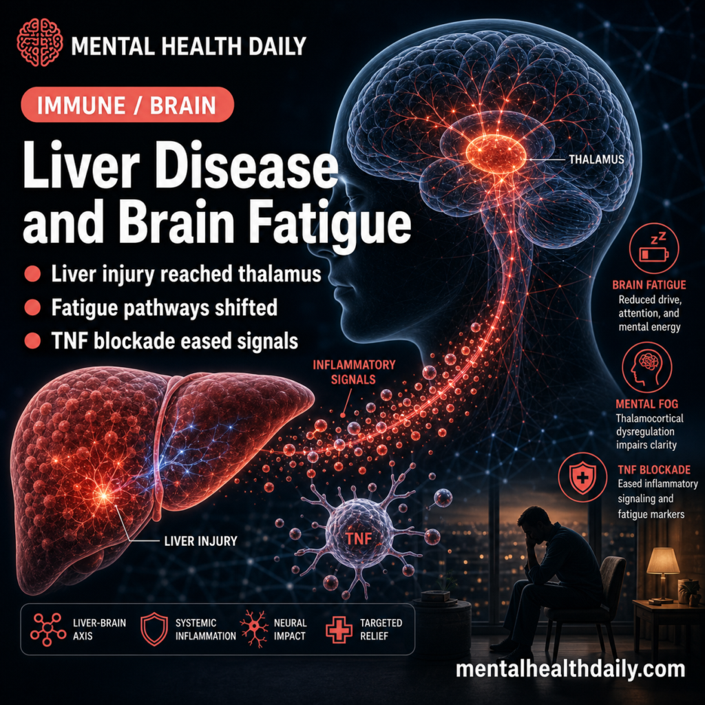
A 2026 mouse study linked cholestatic liver injury to smaller thalamic volume than control injury-free mice, broad thalamus gene-expression changes, and partial pathway rescue after systemic tumor necrosis factor neutralization.
Research Highlights
- Thalamic volume decreased: bile duct ligation (BDL; a mouse model of obstructive cholestatic liver injury) reduced thalamic volume vs. sham surgery by magnetic resonance imaging (MRI), with p = 0.0074 and n = 10 mice per group.1
- Gene pathways shifted toward lower neural maintenance: bulk RNA sequencing in 9 sham and 9 BDL thalamus samples found changes consistent with inhibited myelination, neural-cell proliferation, neurite growth, neurotransmission, and central nervous system development.
- TNF neutralization partly reversed the pattern: systemic anti-tumor necrosis factor treatment in 4 RNA-seq thalamus samples increased predicted activity for neuron development, neuritogenesis, neurotransmission, synaptic transmission, neuroprotection, and neural-cell proliferation pathways.
- Myelination stayed altered: anti-TNF treatment left thalamic myelination-pathway dysregulation in place, so the intervention looked partial across the 10-day model.
- Evidence strength is mechanistic and preclinical: the 2026 study supports a liver-to-brain inflammatory pathway in mice; fatigue behavior testing and human anti-TNF treatment data remain future steps.
Cholestatic liver disease means bile flow is impaired. Primary biliary cholangitis (PBC; an autoimmune disease that damages small bile ducts) and primary sclerosing cholangitis (PSC; a bile-duct scarring disease) can produce fatigue, cognitive symptoms, low mood, anxiety, sleep disruption, and reduced quality of life even when liver-directed blood markers do not fully explain how patients feel.23
Almishri et al. asked whether liver inflammation can alter the thalamus, a deep brain hub involved in alertness, motivation, attention, sensory integration, and communication between cortical and subcortical systems.4 The researchers used BDL in mice to model cholestatic liver injury, then measured thalamic structure with MRI and thalamic gene-expression patterns with RNA sequencing (RNA-seq; a method for measuring which genes are more or less active in tissue).
BDL Mice Had Smaller Thalamic Volume 10 Days After Surgery
The first result was anatomical. At 10 days after surgery, BDL mice had significantly lower thalamic volume normalized to total brain volume compared with sham-operated controls. The MRI figure reported p = 0.0074 with n = 10 mice per group.1
The thalamus result fits prior human imaging work in primary biliary cholangitis. Mosher et al. reported altered deep-gray-matter functional connectivity in PBC in 2017, and a later PLOS ONE study reported structural and functional MRI changes in interoceptive brain regions in PBC patients.25 Interoception means brain processing of internal body signals, such as visceral sensation, arousal, and fatigue-like body states.
The mouse study supports a mechanistic bridge from cholestatic liver injury to thalamic biology. A liver-injury model produced a measurable thalamic-volume change and a molecular signature in the same region, which gives the human imaging literature a more concrete experimental anchor.
RNA Sequencing Pointed to Lower Myelination, Growth, and Neurotransmission
RNA-seq method: the researchers sequenced thalamic RNA from 9 sham mice and 9 BDL mice. They defined differentially expressed genes using false discovery rate (FDR; a statistical correction for many simultaneous tests) below 0.05, absolute fold change at least 1.2, and maximum group mean at least 1.
Pathway mapping: those gene lists were then analyzed with Ingenuity Pathway Analysis (IPA), a curated pathway tool that maps gene-expression changes onto predicted biological functions.
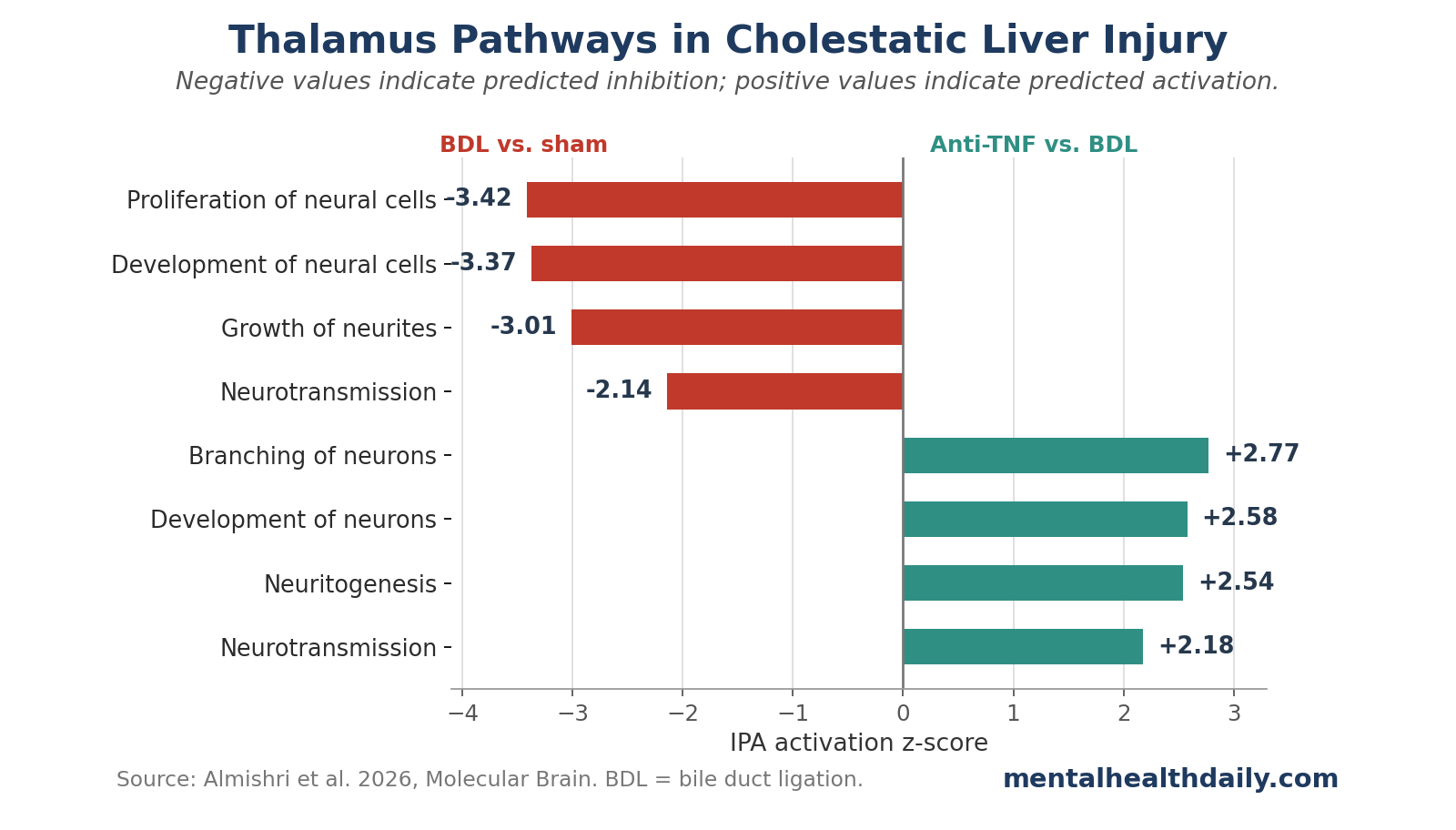
Several thalamic pathways moved in the same direction. The study reported predicted inhibition of:
- Myelination signaling: myelin is the insulating material that helps neural signals travel efficiently; PLP1 messenger RNA, a marker tied to myelin integrity, was lower in BDL mice.
- Neural-cell development: development of neural cells had B-H p = 2.81E-37, predicted decreased activity, activation z-score −3.374, and 343 overlapping molecules.
- Neurite growth: growth of neurites, meaning axons and dendrite-like neural projections, had B-H p = 4.68E-15, predicted decreased activity, z-score −3.013, and 142 molecules.
- Neurotransmission: neurotransmission had B-H p = 8.64E-17, predicted decreased activity, z-score −2.141, and 143 molecules.
- Neural-cell proliferation: proliferation of neural cells had B-H p = 2.21E-23, predicted decreased activity, z-score −3.415, and 227 molecules.
The qRT-PCR validation supported the pathway direction. Quantitative reverse-transcription polymerase chain reaction (qRT-PCR; a targeted method for measuring specific RNA signals) showed lower Ki67, a cell-proliferation marker, and higher Cdkn1a/p21, a cell-cycle inhibitor, in BDL thalamus compared with controls.
That pattern is more specific than “inflammation affects the brain.” It points to structural-maintenance and signaling systems: myelin, neural growth, synaptic communication, and cell survival.
Thalamus Findings Fit a Larger Fatigue Network
Fatigue in chronic liver disease has often been difficult to treat because liver-directed measures do not map neatly onto patient symptoms. D’Mello and Swain described liver-brain inflammatory communication as a plausible route for fatigue and mood symptoms in inflammatory liver disease.6 The 2026 thalamus study gave that idea a region-specific molecular anchor in a cholestatic mouse model.
The thalamus is a reasonable target because it participates in large-scale brain-network coordination. Shine et al. described the human thalamus as a hub for brain-wide information processing, and thalamic abnormalities have been linked to fatigue across conditions such as multiple sclerosis, post-COVID illness, stroke, and mild traumatic brain injury.47
The relevant functions are easy to lose in technical language, so separating them helps:
- Alertness and arousal: thalamic circuits help regulate whether the brain is ready to process incoming information.
- Motivation and reward: thalamic links with striatal and limbic systems can affect effort, drive, and social behavior.
- Sensory integration: thalamic relay and integration functions shape how bodily and sensory signals reach cortical networks.
- Network coordination: thalamic connectivity helps organize cortical and subcortical communication needed for attention and adaptive behavior.
Fatigue-specific behavioral testing was absent from this paper. The connection to fatigue comes from prior human PBC imaging, prior BDL behavior work, and the thalamus literature.
Anti-TNF Treatment Shifted Neurotransmission and Growth Pathways Upward
Tumor necrosis factor (TNF) is an inflammatory cytokine, meaning a signaling protein used by the immune system. The researchers gave BDL mice systemic anti-TNF neutralizing antibody every other day starting 2 days after surgery, while sham controls received phosphate-buffered saline. The antibody schedule covered days 2, 4, 6, and 8 after surgery.
Anti-TNF treatment did not reduce the severity of liver injury in this model, matching earlier work from the same group. That separation between liver injury and brain signaling strengthens the interpretation: if thalamic gene patterns improve without a clear reduction in liver injury severity, systemic TNF may be part of the communication route from inflamed liver to brain.
The RNA-seq comparison used 9 thalamic samples per sham and BDL group, plus 4 thalamic samples from BDL mice receiving anti-TNF for RNA-seq. The pathway analysis found increased predicted activity in several functions when anti-TNF-treated BDL mice were compared with untreated BDL mice:
- Development of neurons: B-H p = 5.46E-10, predicted increased activity, z-score 2.581, and 43 molecules.
- Neuritogenesis: B-H p = 1.03E-09, predicted increased activity, z-score 2.541, and 37 molecules.
- Neurotransmission: B-H p = 3.90E-15, predicted increased activity, z-score 2.179, and 34 molecules.
- Synaptic transmission: B-H p = 7.02E-12, predicted increased activity, z-score 2.129, and 27 molecules.
- Neuroprotection: B-H p = 2.74E-05, predicted increased activity, z-score 2.046, and 8 molecules.
- Branching of neurons: B-H p = 2.20E-04, predicted increased activity, z-score 2.774, and 17 molecules.
qRT-PCR also supported anti-TNF effects on selected genes. The figure caption reported p values of 0.0434, 0.035, 0.041, 0.0336, and 0.0154 for several anti-TNF-responsive gene measures; ADCY1 was not significant (p = 0.4833).
Myelination Did Not Normalize with TNF Neutralization
The anti-TNF result was partial. The researchers reported that TNF neutralization did not prevent dysregulation of thalamic myelination pathways, either by RNA-seq and IPA analysis or by qRT-PCR for PLP1 messenger RNA.
That limitation is biologically important. Myelination affects conduction speed, network synchrony, and the stability of neural communication. Neuroinflammation can damage neurons, synapses, axons, dendritic projections, and myelination processes, while those injury channels may depend on different inflammatory signals.8
The result suggests at least 2 separable processes in cholestatic liver injury:
- TNF-sensitive signaling changes: neurotransmission, synaptic transmission, neurite branching, neuron development, and cell-proliferation pathways shifted favorably after systemic TNF neutralization.
- TNF-resistant myelin changes: myelination-pathway disruption persisted despite anti-TNF treatment, implying additional inflammatory, metabolic, glial, bile-acid, or vascular mechanisms.
This split keeps the interpretation grounded. Anti-TNF selectively altered several thalamic pathways while leaving myelination unresolved.
Evidence Strength and Treatment Implications
Evidence strength: moderate for a preclinical mechanism, low for direct clinical treatment. The study combined MRI, bulk RNA-seq, pathway analysis, anti-TNF manipulation, and qRT-PCR validation. It also built on prior human PBC imaging and prior liver-to-brain inflammation work.256
The limits are equally direct. This was a male-mouse BDL model, and translation to human PBC or PSC requires clinical studies.
Model gap: BDL is a strong model of obstructive cholestatic injury, while human cholestatic disease includes immune, genetic, chronic, sex-specific, and treatment-related features that the model only partially captures.9
Endpoint gap: the study measured thalamic structure and gene pathways; patient fatigue scores, cognitive tests, sleep outcomes, depression scales, and clinical response to TNF inhibitors remain open endpoints.
For now, the treatment implication is narrow and testable: systemic TNF signaling may contribute to thalamic molecular changes during cholestatic liver injury, and future work should test whether blocking that pathway changes behavior, fatigue-like measures, or patient symptoms.
Questions About Cholestatic Liver Disease, Thalamus Changes, and Fatigue
How strong is the liver-to-thalamus fatigue evidence?
The evidence supports a plausible liver-to-thalamus pathway in mice and fits prior human imaging work in primary biliary cholangitis. Direct fatigue-behavior testing is the next needed step.
What does TNF neutralization mean?
TNF neutralization means using an antibody to bind tumor necrosis factor, an inflammatory immune signal, so it cannot signal as strongly. In this mouse study, systemic anti-TNF treatment partly reduced thalamic pathway disruption even though it did not appear to reduce liver injury severity.
Could anti-TNF drugs treat fatigue in primary biliary cholangitis?
The current evidence supports future mechanistic and translational work, ideally including fatigue behavior in animals and carefully designed human studies. TNF inhibitors have infection and immune risks, so clinical testing would need a formal risk-benefit rationale.
Why focus on the thalamus?
The thalamus helps coordinate alertness, attention, motivation, sensory processing, and communication between major brain networks. Those functions overlap with fatigue and cognitive symptoms, and several neurological conditions with fatigue show thalamic structural or connectivity changes.
References
- Thalamic Homeostatic Transcriptomic Signatures Are Altered in a Mouse Model of Cholestatic Liver Injury and Are Mitigated by Systemic TNF Neutralization. Almishri W et al. Molecular Brain. 2026;19:28. doi:10.1186/s13041-026-01302-5
- Primary Biliary Cholangitis Patients Exhibit MRI Changes in Structure and Function of Interoceptive Brain Regions. Mosher V et al. PLOS ONE. 2019;14(2):e0211906. doi:10.1371/journal.pone.0211906
- The Inter-Relationship of Symptom Severity and Quality of Life in 2055 Patients with Primary Biliary Cholangitis. Dyson JK et al. Alimentary Pharmacology & Therapeutics. 2016;44(10):1039-1050. doi:10.1111/apt.13794
- The Impact of the Human Thalamus on Brain-Wide Information Processing. Shine JM et al. Nature Reviews Neuroscience. 2023;24(7):416-430. doi:10.1038/s41583-023-00701-0
- Primary Biliary Cholangitis Alters Functional Connections of the Brain’s Deep Gray Matter. Mosher VAL et al. Clinical and Translational Gastroenterology. 2017;8:e107. doi:10.1038/ctg.2017.34
- Liver-Brain Interactions in Inflammatory Liver Diseases: Implications for Fatigue and Mood Disorders. D’Mello C et al. Brain, Behavior, and Immunity. 2014;35:9-20. doi:10.1016/j.bbi.2013.10.009
- Changes in Thalamic Functional Connectivity in Post-Covid Patients with and without Fatigue. Leitner M et al. NeuroImage. 2024;301:120888. doi:10.1016/j.neuroimage.2024.120888
- The Impact of Neuroinflammation on Neuronal Integrity. Tastan B et al. Immunological Reviews. 2024;327(1):8-32. doi:10.1111/imr.13419
- Bile Duct Ligation in Mice: Induction of Inflammatory Liver Injury and Fibrosis by Obstructive Cholestasis. Tag CG et al. Journal of Visualized Experiments. 2015;(96):52438. doi:10.3791/52438