
A 2026 case-control imaging study found that 23 women with premenstrual dysphoric disorder had higher left DTI-ALPS glymphatic index than 27 healthy controls (p = 0.024), plus wider hypothalamus connectivity across emotion and subcortical networks.1 The result adds a brain-fluid-clearance and stress-network layer to PMDD biology, but the evidence is still small-sample imaging rather than a clinical diagnostic test.
Research Highlights
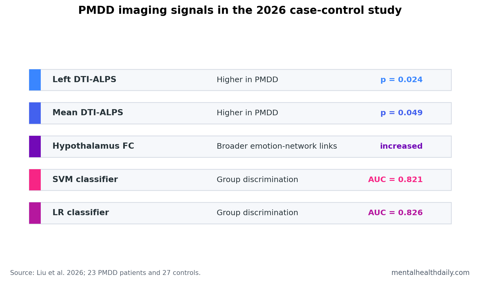
- Glymphatic marker was higher: PMDD patients had higher left DTI-ALPS index than controls (p = 0.024) and higher mean DTI-ALPS index (p = 0.049).
- Hypothalamus connectivity increased vs. controls: the 23-patient group showed higher links with cingulate, frontal, orbitofrontal, insula, temporal, parietal, thalamic, caudate, and lentiform regions.
- Symptoms tracked specific edges: hypothalamus-right inferior temporal connectivity correlated with anxiety scores at r = 0.573 and depression scores at r = 0.480.
- Classifier results were promising but early: support vector machine AUC was 0.821 and logistic regression AUC was 0.826, both with accuracy = 0.780.
- Clinical translation needs restraint: DTI-ALPS is indirect, the sample was 50 people total, and leave-one-out discrimination is not the same as external diagnostic validation.
Premenstrual dysphoric disorder is a cyclical mood disorder in which irritability, depression, anxiety, sleep disruption, and physical symptoms rise in the late luteal phase and impair daily functioning. It is related to menstrual-cycle biology, and the most useful biological question is how hormone-sensitive systems connect with stress, inflammation, sleep, and emotion circuits.
DTI-ALPS means diffusion tensor imaging analysis along the perivascular space. It uses diffusion MRI to estimate water movement along spaces surrounding blood vessels, making it an indirect proxy for glymphatic activity. The glymphatic system is the brain’s fluid-clearance pathway, often discussed in relation to sleep, waste clearance, inflammation, and neurodegeneration.
DTI-ALPS Was Higher in PMDD, Not Lower
Liu et al. scanned women in the late luteal phase, when PMDD symptoms are expected to be active.1 Groups were closely matched by age, menstrual history, progesterone, and estradiol, while PMDD patients had much higher symptom scores: SAS anxiety 51.61 vs. 36.56, SDS depression 56.13 vs. 41.96, DRSP 70.18 vs. 29.22, and PSQI sleep score 8.39 vs. 5.78.
The DTI-ALPS direction is important. Many glymphatic papers frame lower DTI-ALPS as impaired clearance, especially in aging and neurodegeneration. Here, PMDD patients showed higher left DTI-ALPS and higher mean DTI-ALPS. That should be read as altered glymphatic-related diffusion, not automatically as better clearance or worse clearance.
Evidence-strength note: DTI-ALPS is not a direct measurement of cerebrospinal-fluid flow. It is a diffusion-based marker that can be affected by anatomy, motion, vascular factors, and preprocessing. The PMDD finding is a clue about altered physiology, not a stand-alone mechanism.
Hypothalamus Connectivity Reached Stress and Emotion Networks
Hypothalamus functional connectivity describes how closely hypothalamic activity fluctuates with activity in other brain regions during resting-state fMRI. The hypothalamus is central to endocrine, autonomic, stress, sleep, and reproductive signaling, so it is a plausible hub for menstrual-cycle-linked symptoms.
Compared with controls, PMDD patients showed increased hypothalamus connectivity with bilateral anterior/middle cingulate cortex, middle frontal cortex, orbitofrontal cortex, insula, thalamus, caudate, and lentiform nucleus, plus right inferior temporal cortex and left inferior parietal lobe.1
That anatomy is not random. Cingulate and insula regions are often involved in salience, interoception, and emotional distress. Orbitofrontal and middle frontal regions help regulate valuation and cognitive control. Caudate and lentiform structures link motivation, action selection, and affective reactivity.
Symptom Correlations Pointed to Specific Edges
The correlation results make the network finding more interpretable. Right and mean DTI-ALPS indices correlated with hypothalamus-right ACC/MCC connectivity in PMDD patients (r = 0.430 and r = 0.440). Hypothalamus-right inferior temporal connectivity correlated with anxiety and depression scores. Hypothalamus-lentiform nucleus connectivity correlated with DRSP symptom severity.
Plain-English read: the imaging variables were group markers with symptom relevance. Some hypothalamus-linked connections scaled with the symptom burden inside the PMDD group.
Older PMDD imaging work gives that pattern context. Gingnell et al. linked PMDD to altered prefrontal reactivity, and Dan et al. reported altered resting-state network topology in PMDD.2,3 The 2026 study adds hypothalamus-seeded connectivity and DTI-ALPS rather than replacing those emotion-network findings.
AUC Above 0.82 Is Interesting, But Not a Diagnostic Claim
The machine-learning piece used hypothalamus-related connectivity features to distinguish PMDD patients from controls. Support vector machine modeling produced AUC = 0.821 with accuracy = 0.780, sensitivity = 0.739, and specificity = 0.815. Logistic regression produced AUC = 0.826 with accuracy = 0.780, sensitivity = 0.783, and specificity = 0.778.
AUC, or area under the receiver operating characteristic curve, summarizes how well a model separates 2 groups across possible thresholds. AUC above 0.80 is usually worth noticing, but this was leave-one-out validation inside a 50-person sample. External validation in new clinics, scanners, countries, and menstrual-cycle timing windows would be needed before any diagnostic use.
Clinical PMDD diagnosis still depends on prospective symptom tracking across cycles. Imaging may eventually help identify subtypes or mechanisms, but it cannot replace the core clinical pattern: luteal-phase symptoms that remit after menses and impair functioning.
The Glymphatic Link Should Stay Mechanistic
Glymphatic biology is tempting because it connects sleep, inflammation, waste clearance, and neural function. Xie et al. showed in mice that sleep increased metabolite clearance from the brain, making sleep a major glymphatic context.4 PMDD often includes sleep disturbance, so the bridge is plausible.
The bridge still needs direct testing. A stronger PMDD glymphatic study would measure sleep architecture, inflammatory markers, cycle phase, repeated DTI-ALPS across follicular and luteal windows, and symptom change across time. That design could test whether altered glymphatic markers track the symptom cycle or remain stable traits.
For now, the calibrated conclusion is specific: PMDD patients in this small imaging study showed higher DTI-ALPS and stronger hypothalamus connectivity, and several hypothalamus-linked edges tracked symptom scores. That is a credible mechanistic clue, not a clinical endpoint.
Cycle Timing and Sleep Are the Next Hard Tests
The strongest follow-up would scan the same participants at multiple menstrual-cycle phases. PMDD is defined by change across the cycle, so a between-person late-luteal snapshot cannot tell whether DTI-ALPS and hypothalamus connectivity are stable traits, luteal-state markers, or consequences of active symptoms.
Cycle-phase design: repeated follicular and luteal scans would test whether glymphatic and hypothalamus-network signals rise and fall with symptoms. If the signal is present only during symptomatic days, it may index state reactivity. If it persists across phases, it may mark a vulnerability trait.
Sleep measurement: the paper reported higher PSQI sleep scores in PMDD patients, but sleep was not the main mechanistic test. Because glymphatic clearance is strongly tied to sleep physiology, future PMDD studies should include actigraphy, sleep timing, sleep architecture, or at least repeated sleep diaries.
Inflammation and hormones: progesterone and estradiol did not differ between groups in the 2026 sample, but single hormone levels rarely capture cycle-sensitive neurobiology. Repeated hormone measures, inflammatory markers, and symptom ratings would make it easier to test whether connectivity changes are hormone-linked, inflammation-linked, sleep-linked, or all 3.
The careful conclusion is that PMDD imaging is moving beyond generic limbic-reactivity language. Glymphatic and hypothalamus findings give researchers a more specific map, but the map still needs time-resolved validation before it can guide care.
Treatment relevance: the current data do not identify a medication, supplement, device, or behavioral intervention. They suggest measurement targets. If future work confirms the pattern, clinicians might ask whether sleep treatment, circadian stabilization, anti-inflammatory strategies, hormonal interventions, or stress-system treatments shift the same imaging signals.
That is a research program, not a clinical recommendation. For patients now, PMDD diagnosis and treatment remain grounded in prospective symptom tracking, selective serotonin reuptake inhibitors, hormonal strategies when appropriate, sleep care, and individualized risk-benefit decisions.
Validation standard: a stronger imaging study would pre-register follicular and luteal windows, confirm ovulation timing, repeat symptom ratings daily, and test the same classifier in a separate PMDD cohort. That would show whether the DTI-ALPS and hypothalamus-connectivity pattern is reproducible rather than optimized for one small scanner sample.
Replication should also separate sleep disruption from menstrual-cycle phase. If poor sleep drives the glymphatic signal, treatment implications differ from a primarily hormone-sensitive hypothalamus-network mechanism.
External validation should include ordinary clinical heterogeneity: medication exposure, comorbid depression or anxiety, contraceptive use, insomnia severity, and cycle regularity.
That would make the imaging claim sturdier.
The study’s value is therefore anatomical precision. It points to the hypothalamus, cingulate, insula, basal ganglia, sleep-linked clearance biology, and symptom-correlated connections as the next places to test, rather than leaving PMDD inside a vague “hormone mood swings” frame.
Questions About PMDD Glymphatic Imaging
Does higher DTI-ALPS mean PMDD brains clear waste faster?
Not necessarily. DTI-ALPS is an indirect diffusion marker. Higher values show altered perivascular diffusion patterns, but they do not prove faster or healthier glymphatic clearance.
Why focus on the hypothalamus?
The hypothalamus links reproductive hormones, stress physiology, sleep, autonomic regulation, and emotion-related body states. Those systems are central to PMDD symptoms.
Could MRI diagnose PMDD?
No. The classifier results were preliminary and came from 50 people. PMDD diagnosis still requires prospective symptom tracking across menstrual cycles.
References
- Liu C, Xu X, Li Y, et al. Increased glymphatic system activity and hypothalamic connectivity in patients with premenstrual dysphoric disorder. Depression and Anxiety. 2026;2026:3641238. https://doi.org/10.1155/da/3641238
- Gingnell M, et al. Menstrual cycle effects on amygdala reactivity to emotional stimulation in premenstrual dysphoric disorder. European Neuropsychopharmacology. 2013. https://doi.org/10.1016/j.euroneuro.2013.05.003
- Dan R, Canetti L, Keadan T, et al. Resting-state functional connectivity in premenstrual dysphoric disorder. Hormones and Behavior. 2020. https://doi.org/10.1016/j.yhbeh.2020.104782
- Xie L, Kang H, Xu Q, et al. Sleep drives metabolite clearance from the adult brain. Science. 2013. https://doi.org/10.1126/science.1241224