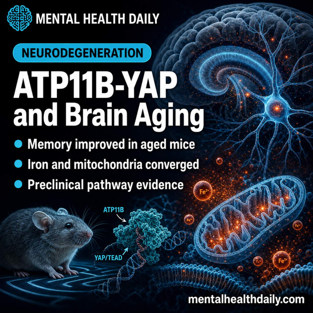
A 2026 mouse and cell study found that ATP11B loss pushed aged brains toward hippocampal iron accumulation, mitochondrial dysfunction, neuronal ferroptosis, and worse memory behavior than wild-type comparison mice; ATP11B overexpression then improved multiple learning and memory tests in 18-month-old mice.
Research Highlights
- Memory signal: ATP11B-deficient mice performed worse than wild-type mice on Morris water maze, Y-maze, and novel-object recognition measures, with behavioral panels using n = 10 per group.
- Iron pathway: Aged and ATP11B-deficient brains showed higher Fe2+ signal alongside blood-brain barrier marker loss, connecting barrier leakage with hippocampal neuron stress.
- Cell map: Single-cell and spatial transcriptomic analyses resolved 7 main cell types and 5 ependymal-cell subtypes, pointing to altered iron transport near the hippocampus.
- Mechanism: ATP11B loss disrupted mitochondrial respiration and quality control, while lactate-linked histone lactylation fed YAP/TEAD activation of ferroptosis and aging genes.
- Evidence limit: ATP11B overexpression improved aged-mouse behavior and dendritic complexity, but this is still preclinical pathway evidence, not a dementia treatment.
ATP11B is a phospholipid flippase, meaning an enzyme that helps keep membrane lipids arranged on the correct side of a cell membrane. In neurons and barrier cells, that membrane-ordering job can touch several aging pathways at once: iron movement, mitochondrial stress, lipid peroxidation, synaptic structure, and cell-death sensitivity.
Ferroptosis is iron-dependent cell death driven by lipid peroxidation, a chemical chain reaction in which iron and reactive oxygen species damage membrane fats. Qi et al. did not show that ATP11B is a ready clinical target in people. The stronger read is narrower and more useful: ATP11B sits at a junction where barrier iron transport, mitochondrial failure, and age-linked memory behavior converge.
ATP11B Loss Made Aged Mouse Brains Look More Iron-Stressed
The study started from a familiar aging-brain problem: the blood-brain barrier, the tightly regulated vascular wall that normally limits what moves from blood into brain tissue, becomes leakier with age. In the hippocampus and cortex of 18-month-old mice, Qi et al. reported lower expression of barrier-integrity markers including Claudin-5, Occludin, and ZO-1 compared with 3-month-old mice.
That barrier change was paired with higher brain Fe2+ signal. Fe2+ is the reduced form of iron that can participate in the Fenton reaction, a chemistry route that generates highly reactive radicals. The article’s mechanistic claim depends on that sequence: barrier vulnerability first changes iron handling, and iron then helps push neurons toward oxidative lipid damage.
The ATP11B angle came from prioritizing transport-related genes. The researchers integrated 3 kinds of evidence: gene-expression correlation with blood-brain substance transport, age-related differentially expressed genes in brain endothelial cells, and genes encoding transport-related proteins. ATP11B emerged among the high-priority candidates, along with genes such as ABCG1, CD9, BOK, and ICAM1.
Behavioral testing: in Atp11b knockout mice, the study reported abnormal social preference, shorter target-zone time in the Morris water maze, reduced novel-arm measures in the Y-maze, and poorer novel-object exploration.
The main memory panels used n = 10 per group, while the survival comparison used n = 16 per group. The PDF reports significance using star thresholds from P < 0.05 through P < 0.0001 rather than extractable effect-size tables.
That reporting style makes the direction clear but limits precision for readers. The study shows that ATP11B loss moved multiple behavioral assays in the same unfavorable direction, but it does not give a clean clinical-sized effect estimate that can be translated into human memory decline.
Single-Cell Mapping Put Ependymal Cells Between Iron and Hippocampal Neurons
Ependymal cells line fluid-filled spaces in the brain and help regulate cerebrospinal fluid composition. They are not the first cells most readers think of in memory decline, but they can influence the chemical environment that neurons sit in.
Qi et al. used single-nucleus RNA sequencing, single-cell RNA sequencing, and spatial transcriptomics to map how Atp11b loss changed brain-cell populations. The single-cell analysis identified 7 main cell types. Within ependymal cells, the researchers identified 5 subtypes, labeled EP0 through EP4.
Ependymal subtype shift: altered ependymal groups were enriched for ion-transport and respiratory pathways. EP0 and EP1 cells decreased in Atp11b-deficient mice, while EP2, EP3, and EP4 cells increased.
In ordinary language, ATP11B loss changed the surrounding transport system that could deliver abnormal iron pressure toward hippocampal neurons.
A compact way to read the cell-mapping evidence:
- Barrier layer: aging reduced hippocampal and cortical tight-junction markers.
- Transport layer: ependymal-cell subtype balance shifted toward ion-transport phenotypes.
- Neuron layer: hippocampal and cortical neuron counts and memory-linked behavior moved in the wrong direction.
Mitochondrial Failure Connected ATP11B to Ferroptosis
Mitochondria generate most cellular ATP, the energy currency neurons use to maintain ion gradients, synaptic signaling, and repair. The 2026 study linked ATP11B loss to mitochondrial respiratory-chain disruption, including altered expression of mitochondrial complex genes and lower complex I activity in cell experiments.
Mitochondrial quality control also shifted. ATP11B knockdown increased fission-related markers such as DRP1 and FIS1 and reduced fusion markers such as MFN1 and MFN2. Fission divides mitochondria; fusion merges mitochondrial networks. Some fission is normal cleanup, but excessive fission plus impaired respiration is a stress pattern rather than healthy renewal.
Mitophagy, the process cells use to remove damaged mitochondria, also increased. Markers including PARKIN, PINK, LC3, OPTN, and p-Ub(S65) rose under ATP11B-deficient conditions. The practical interpretation is not that mitophagy itself is harmful. It is that neurons appeared to be compensating for damaged mitochondria, higher reactive oxygen species, and a growing ferroptosis-prone state.
Epigenetic layer: ATP11B loss increased lactate-linked histone lactylation, a chemical mark on histone proteins that can change gene transcription.
Through the TEAD-YAP complex, that lactylation was tied to genes including Acsl4, Trp53, and Cdkn1a. Acsl4 is especially relevant because it helps load polyunsaturated fatty acids into membrane phospholipids, making cells more vulnerable to ferroptotic lipid peroxidation.

ATP11B Overexpression Improved Memory Tests, But Only in Mice
The rescue experiment is the reason this paper is more than a correlation map. The researchers injected 18-month-old mice with a vector designed to overexpress Atp11b, then tested behavior 1 month later against aged controls and 6-month-old wild-type controls. The main rescue panels again used n = 10 per group.
ATP11B-overexpressing mice had better stride length, longer treadmill time to exhaustion, better balance-beam performance, higher open-field activity, more novel-arm time in the Y-maze, more zone crossings in the Morris water maze, and improved response latency across water-maze training days. Brain tissue then showed greater dendritic length and spine complexity, along with biochemical evidence of ferroptosis rescue and improved mitochondrial fission markers.
Evidence-strength note: this is an animal and cell-model result. It can identify a plausible mechanism and show that manipulating ATP11B changes aging-like behavior in mice. It cannot tell a patient whether ATP11B can be raised safely, whether a human dementia subtype is ATP11B-dependent, or whether an ATP11B-targeted drug would improve cognition.
Rescue logic: the overexpression arm tested reversibility. A purely knockout-based paper could show that ATP11B loss is harmful without showing that restoring the pathway changes outcomes after aging biology is already present.
Qi et al. added that rescue step, which makes ATP11B a stronger mechanistic candidate while still leaving drug delivery, dose, cell specificity, and human relevance unanswered.
Those unanswered pieces are not minor implementation details. A membrane-flippase pathway could affect many tissues, so any future intervention would need evidence that brain-targeted benefit outweighs off-target lipid-transport disruption.
Prior ATP11B Papers Point to Vascular, Synaptic, and Alzheimer Biology
Adjacent ATP11B papers make the 2026 result less isolated:
- Vascular ATP11B: Quick et al. linked loss of heterogeneous ATP11B expression to cerebral small-vessel disease in a rat model, placing ATP11B near vascular brain integrity.
- Synaptic ATP11B: Wang et al. reported that ATP11B deficiency impaired hippocampal synaptic plasticity, which fits the memory-behavior direction in the Qi paper.
- Alzheimer-pathology ATP11B: Zhang et al. tied ATP11B to microglial lipid metabolism and Alzheimer-type pathology, extending the pathway into immune-lipid handling in neurodegeneration.
Ferroptosis literature also fits the direction. Stockwell’s 2022 review framed ferroptosis as an iron-dependent lipid-peroxidation pathway with therapeutic relevance across disease models. Tian et al. connected hippocampal ferroptosis inhibition with improved memory decline in an ovariectomy model. Qi et al. added a more specific route: ATP11B loss changed iron transport, mitochondrial respiration, and lactate/YAP-linked transcription in the same aging-brain model.
Calibrated read: ATP11B remains a preclinical pathway node in age-related cognitive decline. Several independent layers point the same way: barrier leakage, Fe2+ accumulation, altered ependymal transport states, mitochondrial stress, ferroptosis markers, and rescue behavior after ATP11B overexpression.
Questions About ATP11B, Ferroptosis, and Cognitive Decline
Does this mean ATP11B can treat Alzheimer’s disease?
No. The study involved mice, neuronal cells, transcriptomics, and ATP11B overexpression experiments. It supports a brain-aging mechanism, not an Alzheimer’s treatment claim.
Why is iron central to the result?
Iron can be useful in controlled biology, but excess Fe2+ can drive radical chemistry and lipid peroxidation. In this model, ATP11B loss was tied to abnormal iron movement toward hippocampal neurons and ferroptosis-prone mitochondrial stress.
What would make the finding clinically stronger?
Human data would need to show that ATP11B expression, ATP11B-regulated lipid handling, or downstream ferroptosis markers track cognitive decline in people. Drug work would then need to show a safe way to alter the pathway without disrupting essential membrane biology.
References
- Qi W, et al. Targeting ATP11B-YAP axis repairs mitochondrial function and inhibits neuronal ferroptosis to attenuate age-related cognitive decline. Signal Transduction and Targeted Therapy. 2026;11:141. doi:10.1038/s41392-026-02652-1
- Quick S, et al. Loss of the heterogeneous expression of flippase ATP11B leads to cerebral small vessel disease in a normotensive rat model. Acta Neuropathologica. 2022;144:283-303. PubMed
- Wang J, et al. ATP11B deficiency leads to impairment of hippocampal synaptic plasticity. Journal of Molecular Cell Biology. 2019;11:688-702. PubMed
- Zhang Y, et al. ATP11B modulates microglial lipid metabolism and alleviates Alzheimer’s disease pathology. MedComm. 2025;6:e70139. PubMed
- Stockwell BR. Ferroptosis turns 10: emerging mechanisms, physiological functions, and therapeutic applications. Cell. 2022;185:2401-2421. doi:10.1016/j.cell.2022.06.003
- Tian Y, et al. 17 beta-estradiol inhibits ferroptosis in the hippocampus by upregulating DHODH and further improves memory decline after ovariectomy. Redox Biology. 2023;62:102708. doi:10.1016/j.redox.2023.102708