A 2026 Acta Neuropathologica study found that oligomeric tau was linked to early nuclear lamina disruption in Alzheimer’s disease tissue, PS19 tauopathy mice, and human induced pluripotent stem cell (iPSC)-derived neurons.1 The strongest claim is mechanistic: tau oligomers appeared to deform the nuclear envelope and disturb chromatin before overt neurodegeneration, but the work does not yet prove a treatment or clinical biomarker.
Research Highlights
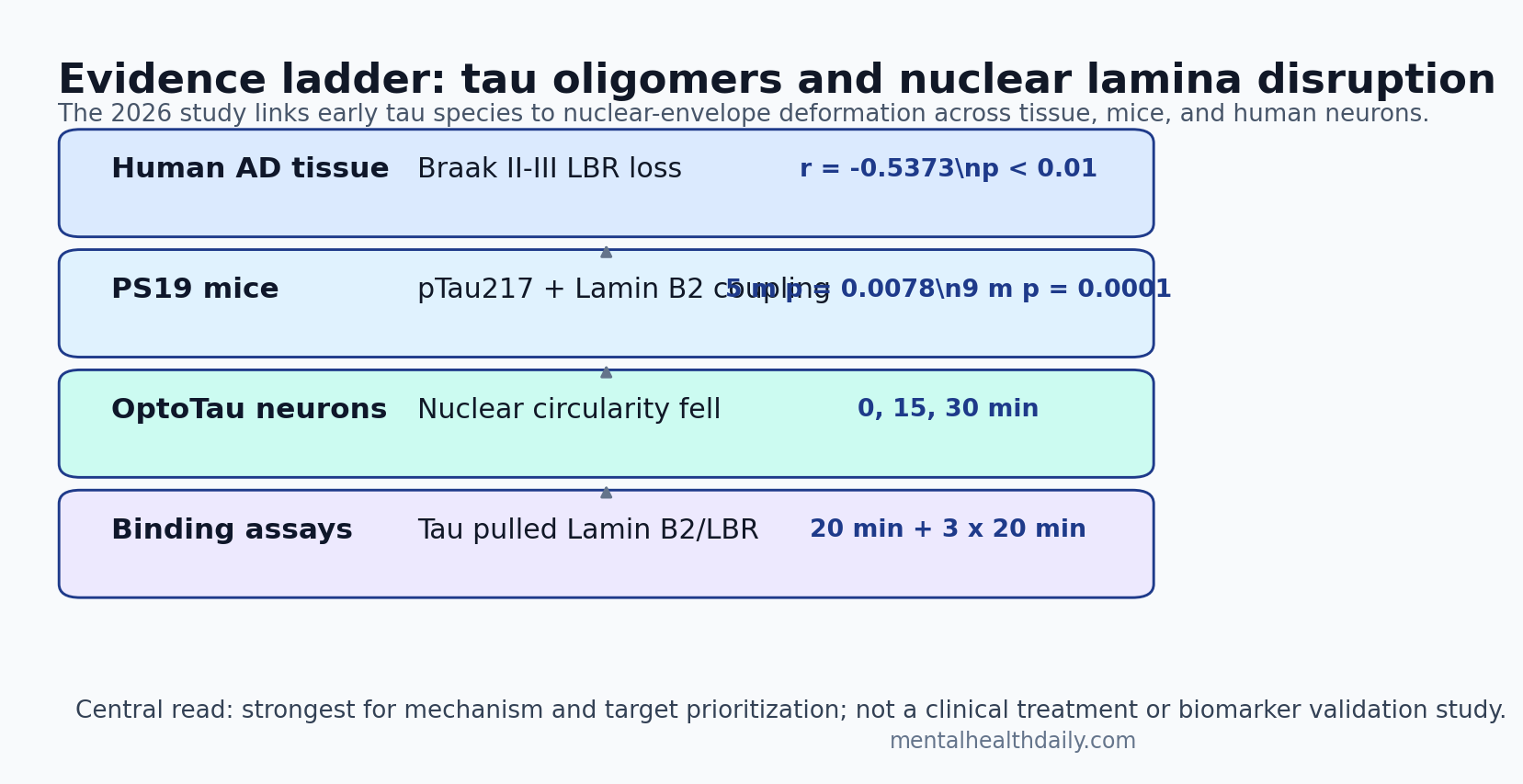
- Lamina damage appeared early: Lamin B receptor nuclear integrity was significantly reduced as early as Braak II-III tissue, and MC1-positive tau pathology correlated with lower LBR integrity (r = -0.5373, p < 0.01).1
- Age did not explain the signal: LBR reduction did not differ by age group (p = 0.5710), while Alzheimer diagnosis remained linked to lower LBR levels (p = 0.0019).1
- PS19 mice showed the same direction: Nuclear pTau217 and internal Lamin B2 were significantly coupled in 5-month and 9-month PS19 mice (p = 0.0078 and p = 0.0001), not in wild-type controls.1
- OptoTau deformed nuclei quickly: Human iPSC-derived neurons showed declining nuclear circularity across 0, 15, and 30 min after light-induced OptoTau oligomerization, while mCherry controls stayed stable.1
- Binding assays supported contact: Co-immunoprecipitation after 20 min and 3 x 20 min light protocols pulled Lamin B2 and LBR with tau complexes, supporting a close tau-lamina interaction.1
Tau oligomers are small aggregated tau assemblies that appear before mature neurofibrillary tangles. In this paper, the reader-facing shift is from “tau tangles accumulate late” to “early tau assemblies may physically stress the nucleus.”
Nuclear lamina means the protein scaffold lining the inside of the nuclear envelope. It helps preserve nuclear shape, anchors chromatin, and supports normal gene regulation. If tau distorts that scaffold, Alzheimer’s disease biology also becomes a genome-organization problem.
Braak II-III Tissue Already Showed Lamin B Receptor Loss
Yuan et al. examined post-mortem Brodmann area 10 prefrontal cortex tissue across Braak stages. Braak staging is a neuropathology scale that tracks the spread of tau pathology through the brain. The researchers stained for MC1, a misfolded tau marker, and Lamin B receptor (LBR), a nuclear-envelope protein that helps tether heterochromatin to the nuclear edge.1
Asymptomatic Braak I-III tissue showed relatively continuous LBR staining and minimal MC1 signal. By Braak IV, LBR staining became fragmented near early MC1 aggregates. Braak V-VI tissue showed more severe LBR disruption and heavier tau pathology.1
The important quantitative detail is timing. LBR nuclear integrity decreased significantly as early as Braak II-III, before the large late-stage tangle burden dominated the tissue. MC1-positive tau pathology correlated with lower LBR integrity (r = -0.5373, p < 0.01).1
Age was an obvious confounder because older brains can show nuclear-envelope changes. The study directly checked that issue: LBR reduction did not differ significantly across age strata of 59-74, 75-90, and >90 years (p = 0.5710), while Alzheimer cases had significantly lower LBR levels than controls (p = 0.0019).1
PS19 Mice Linked pTau217 to Nuclear Invagination
PS19 mice carry a human P301S MAPT mutation and develop tauopathy features including phosphorylated tau, neurofibrillary-tangle-like inclusions, neuronal loss, and brain atrophy. Yuan et al. used them to ask whether tau pathology alone could reproduce the nuclear-lamina pattern seen in human Alzheimer tissue.1
The researchers classified nuclei as invaginated when internal Lamin B2 was at least 0.3 of total Lamin B2. That threshold matters because it turns a visual impression into a measurable nuclear-folding endpoint.1
Compared with wild-type mice, 5-month and 9-month PS19 mice showed more phosphorylated tau and more misshapen nuclei. Nuclear pTau217 intensity correlated with internal Lamin B2 only in PS19 mice, with significantly nonzero slopes at 5 months (p = 0.0078) and 9 months (p = 0.0001). The difference in slopes across groups was also significant (p = 0.012).1
Electron microscopy added structural detail. Six-month and 9-month PS19 mice showed nuclear envelope invaginations and chromatin loosening compared with age-matched controls. The chromatin pattern was strongest near the nuclear membrane zone, the outermost 20% of the nucleus where lamina-associated domains normally help anchor heterochromatin.1
Interpretive key: the mouse evidence ties tau pathology to nuclear shape and chromatin organization alongside classic cytoplasmic tangle pathology.
OptoTau Tested Whether Oligomeric Tau Could Deform Nuclei Directly
The human iPSC-neuron experiment is the causal center of the paper. OptoTau is a light-inducible tau construct: blue light drives a Cry2Olig-linked 4R1N tau protein to oligomerize, letting researchers watch tau aggregation unfold on a controlled time course. The mCherry control uses the same light-responsive system without tau.1
Across 0, 15, and 30 min of blue-light exposure, OptoTau neurons showed a significant decline in nuclear circularity, meaning nuclei became less round and more deformed. mCherry controls remained stable. Particle tracking showed OptoTau aggregates converging toward nuclear cavities and crossing the nuclear boundary, while control granules moved more randomly.1
A longer 60-min protocol increased disease-relevant tau markers including TOMA2 oligomeric tau, pTau217, and MC1 misfolded tau. TUJ1, a beta-III tubulin neuronal marker, declined under OptoTau conditions, suggesting cytoskeletal stress or neuronal injury in the model.1
Co-IP Pulled Lamin B2 and LBR With Tau Complexes
Visual overlap can mislead, so the binding assays matter. Co-immunoprecipitation is a method that pulls down one protein or protein complex and then tests what comes along with it. Yuan et al. used an anti-mCherry pull-down and a TOMA2 oligomeric-tau pull-down to test whether tau complexes physically associated with nuclear lamina proteins.1
After three 20-min light cycles, Lamin B2 and LBR co-precipitated with tau-containing complexes. A separate 20-min exposure helped show that mCherry-Cry2Olig alone did not explain the tau phosphorylation signal, while OptoTau did. The TOMA2 pull-down enriched higher-molecular-weight tau species above 150 kDa and pulled Lamin B2 and LBR under light-induced conditions.1
Translation-stress boundary: the paper measured puromycin labeling through the SUnSET method, a nonradioactive way to estimate new protein synthesis. Protein-translation signals changed with construct and oligomerization state, but gene-expression dysregulation remained a plausible downstream pathway rather than a full transcriptomic result in this study.1
Earlier Tau Nuclear Papers Pointed in the Same Direction
The Yuan study does not stand alone. Frost et al. reported in 2016 that lamin dysfunction mediated neurodegeneration in tauopathy models, placing the nuclear lamina inside the tau toxicity pathway rather than outside it.2 Paonessa et al. later showed that tau-mediated frontotemporal dementia models could deform the nuclear membrane and disrupt nucleocytoplasmic transport, the movement of molecules between nucleus and cytoplasm.3
Chromatin evidence also predates this paper. Frost et al. reported in 2014 that tau promoted global chromatin relaxation, a shift from tightly packed heterochromatin toward a more open and potentially dysregulated state.4 Eftekharzadeh et al. connected tau to nuclear transport disruption in Alzheimer’s disease.5
Yuan et al. sharpened that literature in 3 ways:
- Human staging: LBR disruption appeared early across Braak stages and was not explained by age strata.
- Model convergence: human tissue, PS19 mice, electron microscopy, live iPSC-neuron imaging, and co-IP assays all pointed toward tau-lamina interaction.
- Oligomer focus: induced early tau oligomerization was enough to deform nuclei within minutes in human neurons.
Clinical Translation Is Target Validation, Not Treatment Evidence
The clinical temptation is to treat nuclear lamina stabilization as the next Alzheimer therapy. The data are not there yet. This is a disease-mechanism paper, not a drug trial, diagnostic validation study, or longitudinal biomarker study in living patients.
Evidence-strength note: the human evidence is post-mortem and cross-sectional by Braak stage. The causal evidence comes from PS19 mice and an engineered OptoTau iPSC-neuron system. Those layers make the mechanism credible, but they cannot show that blocking tau-lamina binding slows cognitive decline in people.
The near-term value is prioritization. If tau oligomers damage neurons partly by binding Lamin B2/LBR, deforming the nuclear membrane, and loosening chromatin, then drug screens can ask more specific questions than “does this reduce tau?” They can ask whether a compound prevents tau-lamina binding, preserves nuclear circularity, keeps lamina-associated chromatin organized, or normalizes protein-translation stress.
That target logic also creates safety constraints. Nuclear lamina proteins are not optional decoration; they help organize the genome. A therapy aimed at nuclear stability would need cell-type specificity, timing, reversibility, and evidence that it preserves normal nuclear architecture rather than freezing damaged cells in a different abnormal state.
Questions About Tau Oligomers and Nuclear Lamina
Does this prove Alzheimer’s disease is a nuclear membrane disease?
No. The paper supports a nuclear-lamina mechanism inside Alzheimer and tauopathy biology. Alzheimer’s disease still includes amyloid, tau, inflammation, synaptic failure, vascular factors, aging biology, and clinical heterogeneity.
Why focus on tau oligomers instead of tangles?
Oligomers appear earlier than mature tangles and may be more mobile, making them plausible drivers of early cellular stress. Yuan et al. directly induced oligomerization and observed nuclear deformation within 30 min.1
Could Lamin B receptor become an Alzheimer biomarker?
Possibly, but not from this paper alone. The study used post-mortem tissue and model systems. A biomarker claim would need living-patient assays, longitudinal staging, specificity checks, and evidence that the measure predicts clinical progression.
How should researchers test the tau-Lamin B2/LBR mechanism next?
A strong next step would test whether preventing tau-Lamin B2/LBR interaction preserves nuclear shape, chromatin organization, and neuronal survival without harming normal nuclear function.
References
- Yuan S, Essepian N, Roberts R, Sherman E, Wang Q, Erisir A, et al. Tau oligomerization induces nuclear lamina invagination and chromatin remodeling in Alzheimer’s disease. Acta Neuropathologica. 2026;151:43. doi:10.1007/s00401-026-03018-1
- Frost B, Bardai FH, Feany MB. Lamin dysfunction mediates neurodegeneration in tauopathies. Current Biology. 2016;26:129-136. doi:10.1016/j.cub.2015.11.039
- Paonessa F, Evans LD, Solanki R, Larrieu D, Wray S, Hardy J, et al. Microtubules deform the nuclear membrane and disrupt nucleocytoplasmic transport in Tau-mediated frontotemporal dementia. Cell Reports. 2019;26:582-593.e5. doi:10.1016/j.celrep.2018.12.085
- Frost B, Hemberg M, Lewis J, Feany MB. Tau promotes neurodegeneration through global chromatin relaxation. Nature Neuroscience. 2014;17:357-366. doi:10.1038/nn.3639
- Eftekharzadeh B, Daigle JG, Kapinos LE, Coyne A, Schiantarelli J, Carlomagno Y, et al. Tau protein disrupts nucleocytoplasmic transport in Alzheimer’s disease. Neuron. 2018;99:925-940.e7. doi:10.1016/j.neuron.2018.07.039