hit counter

Group fMRI Misread Cognitive Control in 4,423 Youth

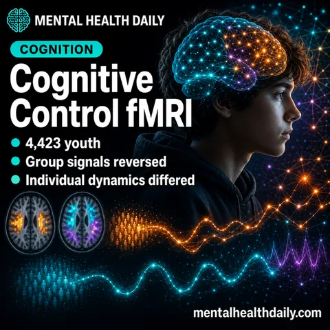
MHD featured image for group fMRI and individual cognitive-control brain signals.

A 2026 ABCD stop-signal fMRI study found that group-level cognitive-control brain signals often failed to describe within-person brain dynamics: across 16 comparisons, between-person and within-person associations diverged, and several reversed direction.1 The practical warning is narrow but sharp: a brain pattern that separates people in a group may be the wrong map for explaining how …

Read more

Language and Theory of Mind Separate by Age 3 in Child Brain Scans

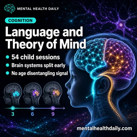
MHD featured image for language and theory of mind brain development in children.

A 2026 fMRI study found language and theory-of-mind activation already separated in 54 child sessions from ages 3-9, with strict child overlap near zero in the left superior temporal lobe (Dice 0.015) and no evidence that the 2 systems disentangled with age.1 The result pushes against a simple developmental story in which language grows out …

Read more

Childhood Anxiety Linked to DNA Methylation and DMN-Limbic Development

MHD featured image for childhood anxiety, DNA methylation, and DMN-limbic network development.

A 2026 Singapore birth-cohort preprint found a conditional childhood-anxiety signal: the cord-blood DNA methylation component predicted less age-13 anxiety only in boys in the low DMN-limbic trajectory group from ages 4.5 to 10.5 years. The paper is useful for mapping developmental risk biology, but it is not a screening test and it does not show …

Read more

Naltrexone Reduced Reappraisal Distress in 38-Person Threat fMRI Trial

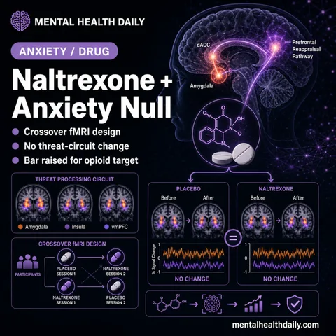
Photoreal illustration of brain with vmPFC, amygdala, and threat-processing circuitry highlighted under pharmacological challenge.

The endogenous opioid system has been a candidate target for novel anxiety treatments, but a 2026 placebo-controlled crossover fMRI study in 38 healthy volunteers ran opposite the hypothesis: instead of increasing threat-related distress and amygdala reactivity, 50 mg oral naltrexone reduced subjective distress during cognitive reappraisal (p = 0.044, d = 0.57) and shifted ventromedial …

Read more

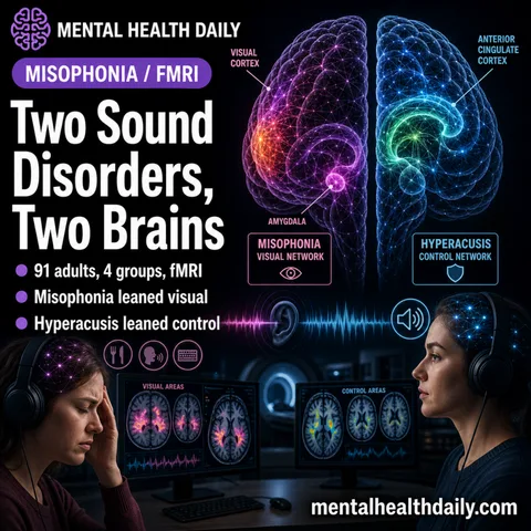
Misophonia vs Hyperacusis fMRI: 91 Adults Show Different Brain Networks

Photoreal illustration of brain regions responding to sound waves with overlapping but distinct activation maps, conveying two sound-sensitivity disorder profiles.

A 2026 task-based fMRI study of 91 young adults separated misophonia from hyperacusis inside the same “sound sensitivity” experiment: misophonia-containing groups showed visual association cortex and ACC-visual network changes during unpleasant sounds, while key hyperacusis contrasts showed weaker salience-control connectivity than misophonia.1 The scan data support separating trigger-specific sound aversion from loudness-driven sound intolerance, while …

Read more

Emotional Memory fMRI Separates Arousal From Valence

Photoreal illustration of brain with amygdala and prefrontal cortex highlighted, conveying dual-pathway emotional memory mechanism.

A 2026 fMRI study of 1,006 healthy young adults found that emotional pictures were remembered better than neutral pictures, but the brain signal split after arousal was modeled: amygdala and insula effects dropped out, while negative and positive valence kept separate cortical encoding patterns.1 Research Highlights Emotional pictures had the recall advantage: in the full …

Read more

Anxious Depression fMRI Meta-Analysis Finds Left Temporal Activation

Editorial brain-map card for anxious depression with highlighted limbic and prefrontal regions from an earlier draft framing.

A 2026 fMRI meta-analysis of 11 studies found a narrow anxious-depression signal: 829 anxious-depression patients differed from 681 non-anxious MDD patients in left middle temporal gyrus activation (SDM-Z = 2.046, p = 0.020), while healthy-control contrasts centered on the anterior commissure and right middle frontal gyrus. The calibrated interpretation is not “amygdala and insula hyperactivity …

Read more