Extra-Virgin Olive Oil Raised Brain Connectivity in a 9-Person Pilot
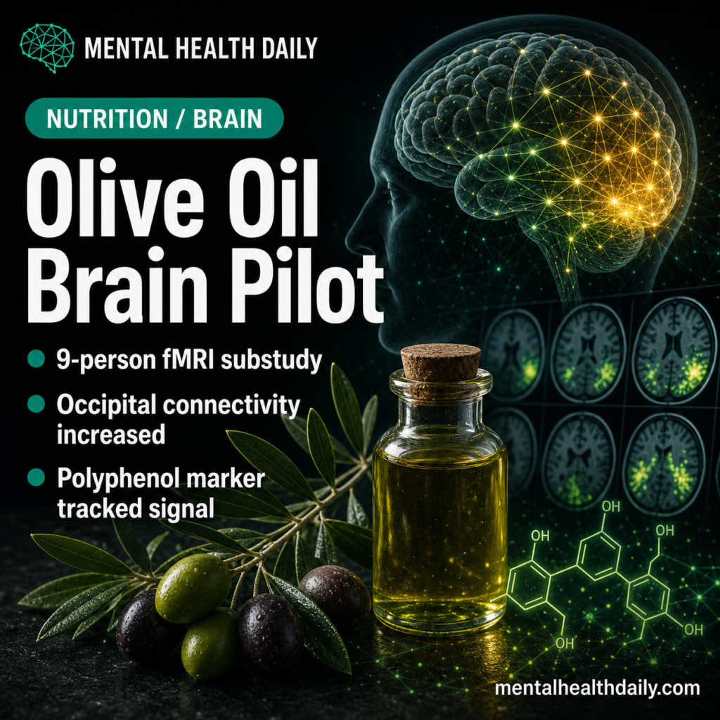
A 9-person randomized crossover neuroimaging substudy found that high-polyphenol extra-virgin olive oil increased occipital resting-state functional connectivity compared with regular olive oil, beta = 0.20, 95% CI 0.03 to 0.37, p = 0.016.1 The result is a measurable brain-signal finding, but the clinical claim stays small: connectivity changed, cognition was not the endpoint. Research Highlights …