
A 2026 automated MRI analysis of 153 adults with first cortical middle cerebral artery stroke found that remote deep gray nuclei changes were already measurable within 21 days, and 2 asymmetry markers independently predicted 3-month disability change: putamen volume Z-score asymmetry (β = −0.24; p = 0.021) and thalamic apparent diffusion coefficient asymmetry (β = 0.24; p = 0.021).1 The result pushes post-stroke prognosis beyond the visible cortical infarct: connected subcortical hubs may carry early recovery information that lesion-volume summaries miss.
Research Highlights
- 153 patients were analyzed: the Lausanne cohort included adults with first acute-subacute cortical MCA stroke, MRI within <21 days, and no infarct directly involving the deep gray nuclei.
- Automated segmentation was feasible: MorphoBox successfully segmented 153 of 169 technically eligible scans after exclusions, or 91% of the initially enrolled cortical MCA stroke cases.
- Stroke-side nuclei had lower Z-scores than opposite-side nuclei: ipsilateral thalamic Z-score difference was 0.15 (p = 0.0011), and ipsilateral caudate Z-score difference was also 0.15 (p = 0.006).
- Thalamic signals split by mechanism: thalamic volume difference tracked infarct volume (β = 0.27; p = 0.001), while thalamic diffusion difference tracked time from stroke onset (F = 6.55; p = 0.0003).
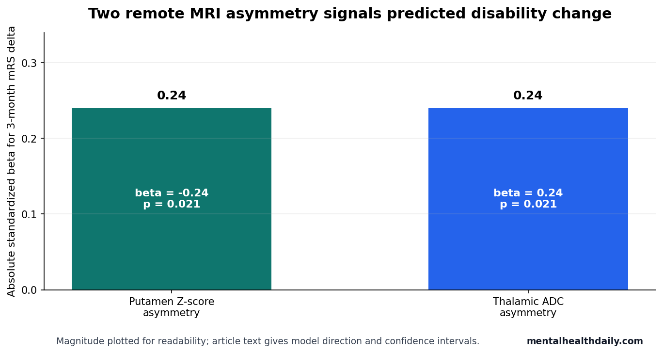
- 3-month disability change had 2 MRI predictors: putamen Z-score asymmetry and thalamic ADC asymmetry independently predicted modified Rankin Scale delta, both with p = 0.021.
Deep gray nuclei are subcortical brain structures such as the thalamus, caudate, putamen, and pallidum. They sit below the cortex but are wired into motor, attention, cognitive-control, and arousal networks that can be affected after a cortical stroke even when the infarct itself does not enter those nuclei.
Modified Rankin Scale (mRS) is a 0-to-6 disability scale used after stroke, where higher values mean more functional dependence or death. In this study, the key outcome was mRS delta: the change between pre-stroke function and 3-month follow-up function.
153 Cortical MCA Stroke Patients Had Remote Subcortical MRI Changes
Zenkhri et al. studied adults admitted to Lausanne University Hospital with first cortical middle cerebral artery (MCA) ischemic stroke between 2018 and 2020. Patients were included if they had 3T MRI with diffusion-weighted imaging and T1-MP-RAGE structural imaging within <21 days of last-known-well time.
The final analyzed sample included 153 patients, mean age 69.9 ± 15.3 years, with 85 men and 68 women. Mean infarct volume was 8.3 ± 20.5 mL, and the mean time from stroke onset to MRI was 56.5 ± 78.5 hours.
MorphoBox is an automated MRI morphometry tool that segments brain structures and compares regional volumes with age- and sex-matched normative ranges. The study used those outputs to calculate deep gray nuclei volume Z-scores, then mapped segmented structures onto apparent diffusion coefficient images to quantify diffusion signal.
Apparent diffusion coefficient (ADC) is an MRI-derived measure of water mobility in tissue. After ischemic injury, ADC can change as cells swell, axons degenerate, myelin breaks down, and tissue structure reorganizes.
Thalamus and Caudate Z-Scores Were Lower on the Stroke Side Than the Opposite Side
The strongest early volume signal was lateralized. Ipsilateral thalamic Z-scores were significantly lower than contralateral thalamic Z-scores, with a contralateral-minus-ipsilateral difference of 0.15 ± 0.57 (p = 0.0011). Caudate Z-scores showed the same mean difference of 0.15 ± 0.65 (p = 0.006).
Putamen and pallidum volume Z-scores did not show the same simple side-to-side difference. Their role became clearer when the researchers used asymmetry indices to predict later disability change.
Plain-language interpretation: the cortical stroke was not physically inside the thalamus or caudate, but those connected nuclei already looked smaller on the stroke side during the acute-subacute window. That pattern fits remote network injury rather than direct infarction of the nuclei themselves.
Infarct Volume and Time Since Stroke Separated the Thalamic Signals
Thalamic volume difference and thalamic diffusion difference did not behave like the same biomarker. The volume Z-score difference between thalami was independently associated with infarct volume (β = 0.27; 95% CI 0.12 to 0.42; p = 0.001). Larger cortical infarcts were linked to a larger thalamic volume imbalance.
Thalamic ADC difference was associated with time from stroke onset to MRI instead (F = 6.55 [3, 148]; p = 0.0003), and thalamic ADC asymmetry showed a similar time relationship (F = 7.04 [3, 148]; p = 0.0002). The diffusion signal looked more like an evolving injury process than a simple reflection of infarct size.
- Volume signal: thalamic Z-score difference tracked infarct burden.
- Diffusion signal: thalamic ADC difference tracked timing after stroke onset.
- Clinical implication: early subcortical MRI changes may carry different information depending on whether the metric is structural volume or water-diffusion behavior.
Putamen and Thalamic Asymmetry Predicted 3-Month Disability Change
Pre-stroke mRS was available for 120 patients, and 3-month mRS was available for 97 patients. In the model predicting 3-month mRS itself, pre-stroke mRS was the dominant clinical predictor (β = 0.58; 95% CI 0.43 to 0.73; p < 0.001), which is expected because baseline disability strongly shapes follow-up disability.
For mRS delta, 2 remote MRI asymmetry markers remained independently associated with functional change. Putamen Z-score asymmetry predicted mRS delta (β = −0.24; 95% CI −0.44 to −0.04; p = 0.021), and thalamic ADC asymmetry also predicted mRS delta (β = 0.24; 95% CI 0.04 to 0.44; p = 0.021).
The putamen result is anatomically plausible because the putamen is part of the basal ganglia circuitry that receives motor and premotor cortical input. The thalamic diffusion result is also plausible because thalamo-cortical pathways can undergo remote degeneration after cortical injury.
Adjacent Stroke Imaging Evidence Points to Remote Network Degeneration
The Zenkhri analysis fits a longer stroke-imaging literature showing that focal infarcts can produce measurable damage in connected regions. Tamura et al. reported thalamic atrophy after MCA territory infarction, and Ogawa et al. used MR imaging to evaluate secondary thalamic degeneration after cerebral infarction in the MCA distribution.2,3
Diffusion work points in the same general direction. Puig et al. found that Wallerian degeneration of the corticospinal tract on diffusion tensor imaging correlated with motor deficit 30 days after MCA ischemic stroke.4 Rosso et al. reported that early ADC changes in motor structures predicted acute-stroke outcome better than lesion volume.5
Other network-level studies broaden the target beyond one tract or nucleus. Duering et al. reported that acute infarcts can cause focal thinning in remote cortex via degeneration of connecting fiber tracts, and Craig et al. linked thalamic diaschisis after perinatal stroke with clinical disability.6,7
Wallerian degeneration means downstream breakdown of axons and myelin after the cell body or upstream connection is injured. Diaschisis means reduced function in a brain region remote from the primary lesion because connected networks have been disrupted.
What This MRI Result Can and Cannot Support
Evidence-strength note: this was a retrospective, single-center observational study. It can show that early deep gray nuclei MRI features were associated with 3-month functional outcome, but it cannot prove that those features cause worse recovery or that the model is ready for bedside prediction.
Several limits matter clinically. Only 97 patients had 3-month mRS data, imaging was measured at 1 time point, and the cohort was drawn from a predominantly Caucasian Swiss hospital population. The study also used mRS rather than detailed cognition, language, gait, or motor-control testing.
The useful next step is external validation: larger multi-center cohorts, repeated MRI time points, lesion-location modeling, and outcome batteries that separate motor recovery from cognitive and psychiatric recovery. If those studies hold, remote deep gray nuclei morphometry could become part of a richer post-stroke recovery profile.
Clinical calibration: this finding is most useful as a risk-enrichment signal, not as a stand-alone forecast. Standard stroke prognosis still starts with age, baseline disability, National Institutes of Health Stroke Scale severity, infarct size, reperfusion status, complications, rehabilitation access, and early functional trajectory. Deep gray nuclei asymmetry would have to improve prediction beyond those ordinary variables before it could change clinical decisions.
Network anatomy: a small cortical infarct can interrupt dense cortico-thalamic, cortico-striatal, or corticospinal pathways. A larger infarct in a less strategically connected region may produce a different recovery pattern.
Automated thalamus, caudate, and putamen measures are an attempt to quantify that network-level injury rather than treating the visible infarct as the whole event.
What would strengthen the signal: a future model should report calibration, discrimination, and decision-curve performance in an outside cohort. It should also test whether the same thalamic ADC and putamen volume-asymmetry features predict specific domains such as gait, arm recovery, language, mood, fatigue, and executive function. Modified Rankin Scale is useful, but it compresses many different recovery problems into one disability score.
For now, the reader-facing interpretation is restrained: early subcortical MRI changes after cortical MCA stroke appear to carry recovery information. The evidence does not yet justify telling an individual patient that a thalamic or putamen asymmetry score determines their 3-month outcome.
Questions About Deep Gray Nuclei MRI After Stroke
Can deep gray nuclei MRI predict recovery for 1 stroke patient?
Not yet. The study found group-level associations in a retrospective cohort. Individual prediction would require external validation, calibration, and comparison with standard clinical stroke predictors.
Why study the thalamus and putamen after a cortical MCA stroke?
Cortical MCA strokes can disrupt cortico-subcortical networks. The thalamus and putamen may change because connected pathways degenerate or reorganize after the primary cortical injury.
Does this finding apply to post-stroke cognition?
The paper measured functional disability with mRS, not detailed cognition. The result is relevant to brain-network recovery, but cognitive prediction needs studies that include neuropsychological outcomes.
References
- Zenkhri S, Marechal B, Corredor-Jerez R, et al. Deep gray nuclei automated assessment in acute-subacute phase of middle cerebral artery cortical stroke helps predict the 3-month outcome. Brain Imaging and Behavior. 2026;20:78. https://doi.org/10.1007/s11682-026-01150-w
- Tamura A, Tahira Y, Nagashima H, et al. Thalamic atrophy following cerebral infarction in the territory of the middle cerebral artery. Stroke. 1991;22(5):615-618. https://doi.org/10.1161/01.str.22.5.615
- Ogawa T, Yoshida Y, Okudera T, Noguchi K, Kado H, Uemura K. Secondary thalamic degeneration after cerebral infarction in the middle cerebral artery distribution: evaluation with MR imaging. Radiology. 1997;204(1):255-262. https://doi.org/10.1148/radiology.204.1.9205256
- Puig J, Pedraza S, Blasco G, et al. Wallerian degeneration in the corticospinal tract evaluated by diffusion tensor imaging correlates with motor deficit 30 days after middle cerebral artery ischemic stroke. American Journal of Neuroradiology. 2010;31(7):1324-1330. https://doi.org/10.3174/ajnr.a2038
- Rosso C, Colliot O, Pires C, et al. Early ADC changes in motor structures predict outcome of acute stroke better than lesion volume. Journal of Neuroradiology. 2011;38(2):105-112. https://doi.org/10.1016/j.neurad.2010.05.001
- Duering M, Righart R, Wollenweber FA, et al. Acute infarcts cause focal thinning in remote cortex via degeneration of connecting fiber tracts. Neurology. 2015;84(16):1685-1692. https://doi.org/10.1212/wnl.0000000000001502
- Craig BT, Carlson HL, Kirton A. Thalamic diaschisis following perinatal stroke is associated with clinical disability. NeuroImage: Clinical. 2019;21:101660. https://doi.org/10.1016/j.nicl.2019.101660