A 2026 Parkinson’s disease mouse study reported that benzhexol hydrochloride induced a measurable hallucination-like behavior state in 6-OHDA parkinsonian mice, and an integrated behavior classifier separated that state from non-PDVH mice with AUC = 0.96 and Youden index = 0.875.1 The careful interpretation is behavioral modeling, not proof that mice had human visual hallucinations.
Research Highlights
- Classifier performance was high: The integrated transition + M1 + M34 behavior model separated induced PDVH-like mice from non-PDVH mice with AUC = 0.96.1
- Optimal discrimination was strong: The same model reached Youden index 0.875, a combined sensitivity-specificity cutoff metric.1
- The drug challenge was specific: Benzhexol hydrochloride was given at 3 mg/kg/day for 14 days after 6-OHDA parkinsonian lesioning.1
- Core behaviors were measurable: M1 lasted 10.64 +/- 2.19 seconds and M34 lasted 7.17 +/- 1.14 seconds, with frequencies of 2.47 and 4.09 per minute.1
- Translation remains limited: A 2026 mouse model can identify behavioral signatures and test pharmacology, but it cannot verify subjective visual experience.
Parkinson’s disease visual hallucinations are false visual perceptions that occur in some people with Parkinson’s disease, especially with advancing disease, cognitive impairment, sleep disruption, and medication complexity. They are clinically important because they predict distress, caregiver burden, and treatment difficulty.
6-OHDA is a toxin used to damage dopamine neurons in animal models of parkinsonism. Benzhexol, also called trihexyphenidyl, is an anticholinergic drug. Anticholinergic drugs can worsen confusion or hallucinations in vulnerable humans, which is why this model used benzhexol as a pharmacological stressor on a parkinsonian background.
Benzhexol Produced a Hunching-Staring-Head-Twitch State
Zhang et al. first created a parkinsonian mouse model using 6-OHDA lesions. They then administered benzhexol hydrochloride at 3 mg/kg/day for 14 days to induce a Parkinson’s visual-hallucination-like state, abbreviated PDVH in the paper.1
The key behavioral state combined hunching posture, prolonged staring, and embedded head twitching. The researchers labeled 2 core movement states as M1 and M34. M1 involved hunching with staring; M34 involved hunching with head-twitching features. These were not ordinary grooming or locomotor changes.
Modeling advantage: hallucination-like states in animals are hard to study because the animal cannot report what it sees. A measurable behavioral signature gives researchers a way to compare drug states, brain activity, and rescue treatments without pretending the subjective report problem is solved.
AUC 0.96 Came From Combining Timing and Movement Features
The most important result was not one behavior alone. Isolated M1, M34, or transition features each classified the induced state well, with AUC values of 0.90, 0.93, and 0.92. Combining transition information with M1 and M34 improved classification to AUC = 0.96.1
AUC means area under the receiver operating characteristic curve. A value of 0.50 is chance classification, while 1.00 is perfect separation. In this model, 0.96 means the integrated behavior pattern almost completely separated benzhexol-induced PDVH-like mice from non-PDVH mice.
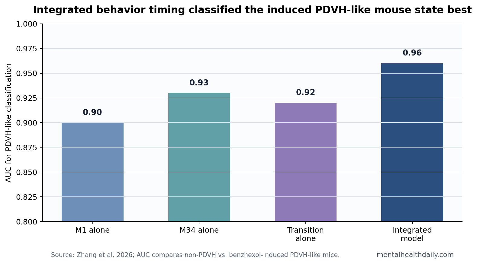
Youden index combines sensitivity and specificity into one cutoff metric. The integrated model reached 0.875, suggesting balanced discrimination rather than one extreme threshold that created many false positives or false negatives.
Pimavanserin Gave the Model a Pharmacology Check
Pimavanserin is a serotonin 5-HT2A inverse agonist approved for hallucinations and delusions associated with Parkinson’s disease psychosis. In a phase 3 trial, Cummings et al. found that pimavanserin improved psychosis symptoms without worsening motor function.2
Zhang et al. used pimavanserin as a validation step. The drug reduced staring and head-twitching durations in the induced mouse model, although not every abnormal behavior fully normalized.1 That partial rescue strengthens the model because a clinically relevant drug moved the expected features in the expected direction.
Panchal and Ondo’s clinical review emphasized the treatment problem in humans: Parkinson’s psychosis management has to reduce hallucinations without worsening parkinsonism, cognition, sleep, or medication side effects.3 A model that can test psychosis-related behavior and motor-state effects together is therefore more valuable than a one-behavior assay.
Anticholinergic Stress Fits Parkinson’s Hallucination Biology
Parkinson’s hallucinations have long been linked to cholinergic dysfunction, visual-processing changes, sleep-wake instability, and medication effects. Anticholinergic drugs can precipitate delirium-like or hallucination-like states because acetylcholine is central to attention, sensory processing, and conscious access.
Volgin et al. reviewed why deliriant and anticholinergic animal models can be informative while still being translationally fragile.4 The Zhang model fits that caution. It is not a full human hallucination model. It is a structured anticholinergic challenge in a parkinsonian animal, with behavior and pharmacology measured in detail.
Best inference: the model gives researchers a repeatable way to quantify a PDVH-like behavioral state. The subjective experience remains unknown, but the timing, posture, transition, and treatment-response data create a more testable system than vague observation.
Human Parkinson’s Hallucinations Are More Than Staring
Human Parkinson’s disease visual hallucinations can range from brief shadows or passage hallucinations to formed people, animals, or scenes. Patients may retain insight early, then lose it as cognition declines or psychosis becomes more persistent. Medication changes can reduce hallucinations in some cases but can also worsen tremor, rigidity, sleep, or mobility.
Clinical mismatch: a mouse state built from hunching, staring, and head twitching cannot reproduce insight, fear, scene content, caregiver distress, or the social consequences of psychosis. Those are human clinical outcomes. The model is strongest when used for mechanistic screening, not when used as a miniature version of the full syndrome.
Mechanistic bridge: the model still has value because cholinergic disruption, serotonergic treatment response, visual-attention changes, and parkinsonian dopamine injury all sit inside the human hallucination literature. A reproducible animal behavior lets researchers test how those systems interact before moving back into patient studies.
The model may be especially useful for separating drug-induced vulnerability from disease-stage vulnerability. Parkinson’s psychosis in patients can emerge from disease progression, sleep disruption, sensory impairment, dementia risk, and medication exposure at the same time. An animal model can hold some of those variables steadier and ask whether an anticholinergic challenge pushes a parkinsonian brain into a distinct behavior state.
Drug-development value: a classifier with AUC 0.96 can screen whether a candidate intervention moves the whole behavior pattern across multiple visible movements. A drug that reduces head twitching while leaving prolonged staring and abnormal transitions intact would be a weaker rescue than a drug that normalizes the integrated signature.
The transition component is especially important. Hallucination-like behavior in a rodent is more convincing when it appears as an organized temporal state rather than as isolated twitch counts. Zhang et al. treated posture, staring, head twitching, and movement transitions as a sequence, which is closer to how clinical behavior is recognized in humans: not one sign in isolation, but a pattern over time.
That temporal framing also reduces false confidence from any single feature. Staring alone can reflect attention, freezing, sedation, or motor impairment. Head twitching alone can reflect serotonergic or motor effects. A combined temporal signature gives the model a more specific behavioral identity.
The same logic applies to treatment testing. A therapy that only suppresses movement could look effective if the model relied on one motor count. A temporal classifier can ask whether the broader PDVH-like state changed, which is a better match for psychosis research.
Safety angle: anticholinergic drugs are already clinically tricky in Parkinson’s disease because they can worsen cognition and hallucinations. The animal model makes that medication-risk axis experimentally visible, even though patient decisions still require human data.
What This Animal Study Can and Cannot Support
Supported: benzhexol can induce a reproducible behavior pattern in 6-OHDA parkinsonian mice; M1, M34, and transition timing classify that state; pimavanserin partly reduces core features.
Not supported: direct evidence that mice saw objects, people, shadows, or scenes. Human visual hallucinations involve perception, attention, memory, insight, and distress. Mouse behavior cannot report those contents.
Evidence-strength note: this is an animal-model paper. Its value is mechanism generation and pharmacology screening. It should not be used to infer prevalence, clinical diagnosis, patient prognosis, or treatment efficacy in humans without clinical evidence.
Best next test: the model becomes more useful if it predicts human-relevant biology rather than only reproducing a visible movement pattern. Stronger follow-up work would pair the behavior classifier with cholinergic markers, visual-attention measures, sleep-wake state, and drug-response profiles that can be compared with Parkinson’s disease cohorts.
Human-pathway mismatch: hallucinations in patients rarely come from one pathway. Lewy pathology, acetylcholine loss, dopamine treatment, impaired vision, sleep disruption, and cognitive decline can all contribute.
The mouse model isolates one medication-risk axis, which is useful for experiments but too narrow for bedside prediction.
Questions About Parkinson’s Visual Hallucination Models
Did the researchers prove mice hallucinated?
No. They created a measurable hallucination-like behavioral state. That is scientifically useful, but subjective visual experience cannot be confirmed in mice.
Why use benzhexol?
Benzhexol is anticholinergic, and anticholinergic burden can worsen hallucinations or confusion in vulnerable humans. Using it in parkinsonian mice creates a biologically plausible stressor for hallucination-like behavior.
Why does AUC matter here?
AUC shows how well behavior features separated induced PDVH-like mice from non-PDVH mice. AUC 0.96 means the combined behavior signature performed much better than chance in this experimental setting.
References
- Zhang Y, et al. Temporal assessment of behavior in Parkinson’s visual hallucination reveals a novel HHS state: hunching with prolonged staring and embedded head twitching. Signal Transduction and Targeted Therapy. 2026;11:146. doi:10.1038/s41392-026-02651-2
- Cummings J, Isaacson S, Mills R, et al. Pimavanserin for patients with Parkinson’s disease psychosis: a randomised, placebo-controlled phase 3 trial. Lancet. 2014;383:533-540. doi:10.1016/s0140-6736(13)62106-6
- Panchal SC, Ondo WG. Treating hallucinations and delusions associated with Parkinson’s disease psychosis. Current Psychiatry Reports. 2018;20:3. doi:10.1007/s11920-018-0869-z
- Volgin AD, Yakovlev OA, Demin KA, et al. Understanding central nervous system effects of deliriant hallucinogenic drugs through experimental animal models. ACS Chemical Neuroscience. 2019;10:143-154. doi:10.1021/acschemneuro.8b00433