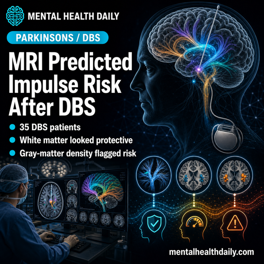
Subthalamic deep brain stimulation (STN-DBS) gives most patients with advanced Parkinson’s disease meaningful motor relief, but a subset develops new or worsened impulse control disorders — pathological gambling, compulsive shopping, hypersexuality, binge eating — while others see preexisting symptoms improve. A 2026 prospective preprint from Loehrer et al. used pre-surgical diffusion MRI to identify microstructural patterns in 35 patients that predicted who would worsen and who would improve after surgery.1
Research Highlights
- Intact frontolimbic white matter predicted improvement in impulsivity from baseline to 6 months after DBS. Higher preoperative fractional anisotropy in the cingulum and tracts adjacent to the insula correlated with larger drops in QUIP-RS score from baseline to 6 months relative to the cohort average.1
- Higher gray-matter neurite density in specific cortical regions predicted worsening relative to the cohort average. Elevated NDI in bilateral paracingulate gyrus, insular cortex, precentral gyrus, right cerebellar lobules I–IV, mesencephalic brainstem, and left putamen correlated with poorer QUIP-RS trajectories from baseline to 6 months — either flat scores or net increases in impulsive-compulsive behavior.1
- Major association tracts mattered. The superior longitudinal fasciculus, uncinate fasciculus, inferior fronto-occipital fasciculus, and forceps minor — tracts connecting frontal control regions to limbic and parietal areas — emerged across multiple diffusion metrics as relevant to ICB risk.1
- The white-matter and gray-matter signatures pointed in different directions. Coherent, well-organized fiber bundles were protective; densely packed neurites in the same anatomical regions in gray matter were vulnerability markers, consistent with separate microstructural roles in the two tissue types.1
- Preliminary — preprint, n = 35. The findings are preregistered hypothesis-generation. Independent replication and prospective validation are needed before diffusion MRI becomes a clinical pre-DBS screening tool.1
Impulse control disorders affect roughly 14–17% of Parkinson’s patients on dopaminergic therapy, with dopamine agonists carrying the highest risk.2 STN-DBS often allows reduction in dopaminergic medication, which can improve impulsivity. But stimulation also spreads through cortico-subcortical networks in ways that can either suppress or amplify impulsive behavior depending on which tracts and circuits are recruited.
Why some patients improve and others worsen is the question this study addresses. The proposed answer: preoperative microstructure of the implicated networks predicts the direction.
Loehrer 2026: 35 Patients, Pre/Post DBS, NODDI Diffusion MRI
The cohort was 35 patients with advanced Parkinson’s disease who met international guidelines for STN-DBS surgery, with full preoperative diffusion MRI and 6-month postoperative clinical follow-up.1
Imaging used:
- Diffusion tensor imaging (DTI): the standard technique for measuring white-matter tract integrity through fractional anisotropy (FA) — how directional the diffusion of water molecules is along axonal bundles. Higher FA usually means more coherent, intact fiber organization.
- Neurite orientation dispersion and density imaging (NODDI): a more advanced model that estimates two separate parameters — neurite density index (NDI), the density of axons and dendrites in a voxel, and orientation dispersion index (ODI), how scattered the neurites are.
The clinical outcome was the Questionnaire for Impulsive-Compulsive Disorders in Parkinson’s Disease-Rating Scale (QUIP-RS), a 4×7 instrument scoring frequency of impulsive thoughts, urges, and behaviors across seven domains (gambling, buying, sexual, eating, punding, hobbyism, and compulsive medication use). Total scores range 0 to 112; subscale scores ≥10 (or ≥7 for punding/hobbyism) define clinically relevant ICDs.
Pre-DBS scans were acquired in the medication-ON state. Six-month follow-up was in the medication-ON / stimulation-ON state, with standardized titration of medications and stimulation parameters.
The analysis correlated voxel-wise diffusion metrics with the change in QUIP-RS score from baseline to 6 months, looking for spatial patterns where preoperative microstructure tracked with postoperative impulsivity outcome.
White-Matter Integrity in the Cingulum and Around the Insula Was Protective
The clearest signal in the white-matter analyses was protective.
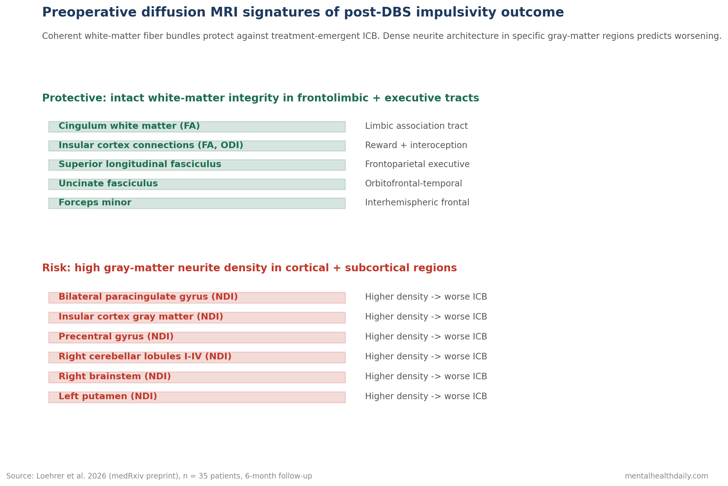
Higher preoperative FA in the cingulum was associated with greater postoperative reductions in impulsive-compulsive symptoms.1 The cingulum is the major limbic association tract connecting anterior cingulate, posterior cingulate, and hippocampal regions — a backbone of emotional regulation and behavioral control circuits.
White matter adjacent to the insula emerged across multiple diffusion metrics, with higher ODI values associating with better ICB outcomes. The insula integrates interoceptive signals with reward processing and emotional valence; intact insular white-matter connectivity supports the regulation of impulsive drives.3
Major association tracts mattered: superior longitudinal fasciculus (frontoparietal executive control), uncinate fasciculus (orbitofrontal-temporal emotional regulation), inferior fronto-occipital fasciculus, and forceps minor (interhemispheric frontal connectivity). Compromised integrity of these tracts predicted worse postoperative outcomes.
The interpretation: when the white-matter scaffolding for inhibitory control and reward regulation is intact preoperatively, STN-DBS plus dopaminergic medication reduction yields net improvement in impulsivity. When it is degraded, the stimulation spreads into a less-regulated network and impulsivity emerges or worsens.
Gray-Matter Neurite Density Pointed the Other Way
The gray-matter findings ran opposite to the white-matter pattern, which is biologically reasonable given that the two tissues serve different roles.
Whole-brain voxel-wise NODDI revealed clusters where higher preoperative NDI was associated with smaller postoperative reductions or even increases in QUIP-RS:1
- Bilateral paracingulate gyrus.
- Insular cortex extending to the uncinate fasciculus.
- Precentral gyrus.
- Right cerebellar lobules I–IV.
- Right mesencephalic brainstem nuclei.
- Left putamen.
The pattern is consistent with reports from other neuropsychiatric conditions involving impaired inhibitory control. NODDI studies in young adult binge drinkers and ADHD have linked higher neurite density in reward-related regions to greater compulsive or impulsive behavior.4,5
The proposed mechanism: dense neurite architecture in cortical territories connected to the STN may produce hyperreactive substrates that amplify the disinhibitory consequences of stimulation through the cortico-basal ganglia-thalamo-cortical loops.
The two patterns — protective coherent white-matter tracts vs. risk-conferring dense gray-matter neurite architecture — reflect the fundamentally different microstructural properties of the two tissues. White-matter signal transmission depends on fiber organization and coherence; gray-matter function depends on the complexity of local neuronal circuitry and the balance of excitation and inhibition.
Post-DBS ICB Drives Financial, Sexual, and Family Disruption
The clinical impact of postoperative impulse control disorders extends well beyond psychiatric symptoms.
Pathological gambling and compulsive shopping can produce financial instability, debt, and family conflict in months. Hypersexuality can disrupt relationships and lead to legally and personally serious consequences. Binge eating contributes to weight gain and metabolic complications.2
For caregivers, ICB onset after DBS is often a more disruptive problem than the motor symptoms the surgery was meant to treat.
Current pre-DBS counseling can warn patients that ICB is a possible outcome but cannot identify which patients are at higher risk. The Loehrer findings, if they replicate, suggest preoperative diffusion MRI could:
- Stratify candidates into higher- and lower-risk categories for ICB development before surgery.
- Improve informed consent by giving individual patients a more concrete risk estimate.
- Guide stimulation parameter choice if certain electrode configurations interact with specific microstructural patterns to produce divergent outcomes.
- Trigger heightened monitoring in higher-risk patients during the first postoperative months when ICB typically emerges.
Pre-DBS Counseling Should Cover Impulse-Control Risk and Family Monitoring
- STN-DBS remains a strong intervention for advanced Parkinson’s motor symptoms. The Loehrer findings do not change the fundamental cost-benefit calculation, which still favors DBS for properly selected patients.
- Preoperative ICB screening is already standard. The QUIP-RS or similar instruments should be administered before surgery, and patients with active or recent ICDs should be flagged for closer postoperative monitoring.
- Diffusion MRI risk stratification is not yet clinical. The findings are exciting but preprint and based on n = 35. Independent replication, ideally with prospective validation in a fresh cohort, is needed before diffusion-derived markers belong in clinical decision-making.
- Postoperative monitoring should include impulsivity. The first 6 to 12 months after DBS surgery are the highest-risk window for ICB emergence. Routine follow-up should ask specifically about gambling, shopping, sexual behavior changes, eating patterns, and compulsive medication use.
- Family involvement matters. Patients are sometimes the last to recognize ICB. Including a family member in postoperative reviews increases detection rate.
35-Patient Diffusion MRI Data Are Not Ready for DBS Screening
It is a preprint. The analysis has not been peer-reviewed at time of writing. Effect sizes, statistical thresholds, and tract-level interpretations may shift in the published version.
The sample is small. 35 patients in a whole-brain voxel-wise diffusion analysis is modest, and the multiple comparison burden across thousands of voxels makes false positives a real concern even with appropriate correction.
The design is correlational. Higher cingulum FA correlates with better postoperative outcome, but the analysis does not establish that intact cingulum integrity causes the better outcome. Confounding by overall brain health, disease stage, or unmeasured cognitive variables remains possible.
NODDI parameters are proxies. NDI estimates neurite density but does not measure functional capacity, neurotransmitter status, or activity-level dynamics. Saying “higher NDI = denser neurites” is a quantitative imaging claim, not a direct biological measurement.
Stimulation parameters were standardized but not personalized. Whether tailored stimulation in patients with at-risk microstructure could prevent ICB emergence is an intervention question this observational design cannot answer.
Questions About DBS, Impulse Control, and Brain Imaging in Parkinson’s
How common are impulse control disorders after STN-DBS?
Estimates vary widely across studies. Roughly 14–17% of Parkinson’s patients on dopaminergic therapy meet criteria for ICDs at any given time, with dopamine agonists carrying the highest baseline risk.2
STN-DBS produces a mixed picture: some patients with preexisting ICDs improve, often via the postoperative reduction in dopaminergic medication; others develop new or worsened impulsive-compulsive behaviors. The Loehrer study tries to identify which preoperative brain features predict each direction.
Which behaviors does QUIP-RS measure?
The Questionnaire for Impulsive-Compulsive Disorders in Parkinson’s Disease covers seven domains: pathological gambling, compulsive buying/shopping, compulsive sexual behavior, compulsive eating, punding (repetitive purposeless activity), hobbyism (excessive engagement in hobbies), and compulsive medication use.1
Each domain is scored 0–4 across four question types (frequency of thoughts, urges, behaviors, and impact), giving a total range of 0 to 112.
Should patients ask for diffusion MRI before DBS surgery?
Not yet. The Loehrer findings are preliminary and have not been replicated or prospectively validated. Standard MRI is already part of pre-DBS workup; adding research-grade NODDI is not yet supported by evidence as clinical risk-stratification.1
If prospective validation confirms the signal, diffusion MRI could become part of standard pre-DBS imaging within several years.
What can be done if ICB emerges after DBS?
Several levers exist. Reducing or stopping the dopamine agonist (if possible without motor decompensation) is often the first step. Stimulation parameter adjustment — reducing voltage or shifting active contacts — can sometimes resolve ICB without sacrificing motor benefit. Behavioral interventions and family support are important adjuncts.2
The Loehrer model could eventually inform which adjustments are most likely to help in a given patient’s microstructural context.
Why does the cerebellum show up in an impulsivity study?
The posterior cerebellum and lobules I–IV connect to prefrontal and limbic networks through cortico-pontine-cerebellar loops. Recent work has expanded the cerebellum’s recognized roles to include cognitive control, affect regulation, and reward processing, alongside its long-known motor coordination function.1
The Loehrer signal in right cerebellar lobules I–IV fits this broader picture and is consistent with cerebellar involvement in the executive aspects of impulse control.
References
- Microstructure predicts impulsive and compulsive behaviour following subthalamic stimulation in Parkinson’s disease. Loehrer PA et al. medRxiv (preprint). 2026. doi:10.64898/2026.04.13.26350763
- Impulse control disorders in Parkinson disease: a cross-sectional study of 3090 patients. Weintraub D et al. Archives of Neurology. 2010;67(5):589-595. doi:10.1001/archneurol.2010.65
- The integrative role of the insula in human cognition and behavior. Uddin LQ et al. Neuron. 2017;95(3):506-520. doi:10.1016/j.neuron.2017.07.025
- NODDI characterizes neurite tissue microstructure in healthy and diseased brains. Zhang H et al. NeuroImage. 2012;61(4):1000-1016. doi:10.1016/j.neuroimage.2012.03.072
- Reward circuitry in addiction. Volkow ND, Wise RA & Baler R. Neuropsychopharmacology Reviews. 2017;42(1):282-292. doi:10.1038/npp.2016.252