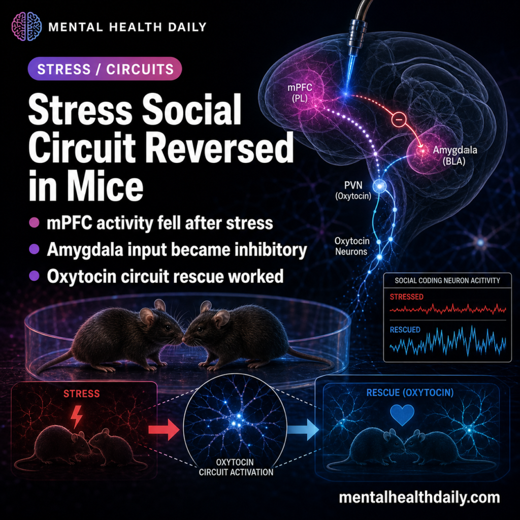
A 2026 mouse circuit study found that restraint stress progressively reduced medial prefrontal social coding: mPFC excitatory-neuron transient rates fell to roughly 70-80% of baseline after acute stress and nearly 50% after chronic stress, while suppressing either basolateral-amygdala input or hypothalamic oxytocin input restored social discrimination and behavior.1 The result is a circuit-specific oxytocin finding, not another simple “oxytocin is prosocial” story.
Research Highlights
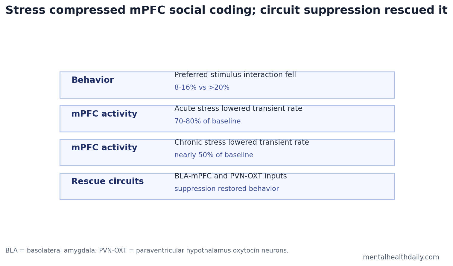
- Stress reduced social engagement: Chronically stressed mice spent only 8-16% of each session interacting with preferred stimuli vs. >20% in controls.1
- mPFC activity fell with stress dose: Calcium imaging recorded 573 baseline neurons, 612 acute-stress neurons, and 683 chronic-stress neurons; transient rates dropped to about 70-80% of baseline after acute stress and nearly 50% after chronic stress.1
- Amygdala input became a rescue target: Suppressing hyperactive BLA-mPFC projections restored mPFC transient rates, pattern decorrelation, and social performance in stressed mice.1
- Oxytocin was state-dependent: Inhibiting PVN-mPFC oxytocin neurons rescued stress-induced social alterations, while suppressing CRH-linked PVN-mPFC neurons did not improve behavior.1
- Clinical translation is distant: These were male mice exposed to 2 h/day restraint across 14 days with viral tools, miniscope imaging, photometry, and optogenetic suppression, not a human treatment study.
Medial prefrontal cortex (mPFC) is a front-brain region that helps represent social context, regulate emotion, and integrate internal state with external cues. In this study, the prelimbic mPFC was the downstream hub where stress-related amygdala and hypothalamic signals converged.1
Oxytocin is a neuropeptide involved in social behavior, stress adaptation, and reproductive physiology. Its effects depend on circuit and state. The same molecule can support social memory in one context and contribute to inhibitory prefrontal imbalance in another.
14 Days of Restraint Stress Progressively Altered Social Testing
Male C57BL/6 mice underwent repeated restraint stress for 2 h/day across 14 days. Researchers tested social preference after acute stress on day 1 and after chronic stress on day 14 using a 3-chamber task. Session 1 compared a conspecific mouse with an object. Session 2 compared a familiar mouse with a novel mouse.1
- Control mice: preferred the mouse over the object and the novel mouse over the familiar mouse.
- Acute-stress mice: retained basic social preference but lost normal novelty preference.
- Chronic-stress mice: lost clear social preference and spent even less time with the novel mouse.
The chronic-stress pattern persisted for at least 2 weeks after stress cessation. Open-field testing did not show generalized hypoactivity, although stressed mice showed anxiety-like reductions in center exploration.1
mPFC Social Coding Dropped From 70-80% to Nearly 50% of Baseline
The study used miniature calcium imaging to monitor mPFC excitatory neurons while mice performed social tests. Recorded populations included 573 neurons at baseline, 612 neurons after acute stress, and 683 neurons after chronic stress.1
Calcium imaging tracks changes in fluorescence from calcium-sensitive proteins, giving a proxy for neuronal activity. Acute stress reduced mPFC transient rates to about 70-80% of baseline; chronic stress pushed responses closer to 50% of baseline. The reduction affected social-stimulus responses and even stimulus-free middle-zone activity.1
Pattern decorrelation means separating neural activity patterns so different stimuli are represented as distinct. Stress compressed those representations, making social vs. nonsocial and familiar vs. novel stimulus coding less separable.
BLA-mPFC Hyperactivity Looked Like Feedforward Inhibition
Basolateral amygdala (BLA) is an amygdala region that links emotionally salient information with prefrontal control systems. Zhang et al. found elevated calcium signals in BLA-mPFC projection neurons during social testing after stress. Activating that projection increased local mPFC GABAergic neuron activity, consistent with feedforward inhibition.1
Suppressing BLA axons in mPFC increased mPFC transient rates during homecage and chamber habituation, restored exploration of social and novel-social stimuli in stressed mice, and improved neural measures of stimulus separation. The rescue was not a generic prosocial boost: inhibiting the BLA-mPFC projection in unstressed controls did not improve novelty preference and reduced general sociability.1
PVN-mPFC Oxytocin Input Became Stress-Linked Inhibitory Drive
Paraventricular hypothalamus (PVN) is a stress and neuroendocrine hub. The study found stress-activated PVN projections to mPFC and then separated peptide-defined circuits. Inhibiting PVN-mPFC oxytocin neurons restored social performance after 1 or 14 days of restraint, while suppressing CRH-linked PVN-mPFC neurons did not improve social behavior.1
PVN-mPFC oxytocin neurons were more active in stressed mice during social testing and increased at restraint onset on days 1 and 14. In mPFC, oxytocin appeared to activate GABAergic inhibitory mechanisms; an oxytocin receptor antagonist improved social novelty preference after acute stress and reduced GABAergic activity.1
Interpretive key: oxytocin was not a universal social-enhancement signal. In the stressed state, this PVN-to-mPFC oxytocin input helped produce inhibitory prefrontal imbalance, and suppressing it rescued social coding.
Two Projection Systems Pointed to the Same Prefrontal Bottleneck
The amygdala and hypothalamic results should be read together. Stress increased upstream inputs that converged on the same medial prefrontal hub and pushed that hub toward weaker stimulus separation. The BLA route supplied threat-salience pressure, while the PVN oxytocin route supplied a stress-linked neuropeptide signal that recruited inhibitory mechanisms inside mPFC.
Converging rescue: if only BLA suppression had worked, the study would look like a standard amygdala-overdrive account. If only PVN oxytocin suppression had worked, the study might be misread as an oxytocin-specific reversal.
The parallel rescues instead point to a prefrontal computation: social behavior deteriorated when mPFC population activity could no longer keep social, nonsocial, familiar, and novel stimuli far enough apart.
Practical translation: the finding argues for circuit-state precision. A molecule or projection can be helpful in one state and harmful in another if it changes which cells dominate the local network. That is why the result should not be flattened into “block oxytocin” or “calm the amygdala.” The actionable scientific claim is narrower: stressed male mice showed excessive BLA-mPFC and PVN-mPFC oxytocin drive, and projection-specific suppression restored mPFC coding.
Stress Models Predict Prefrontal Control Loss
Stress can impair prefrontal cortex function and shift behavior toward more reflexive, amygdala-driven control.2 Zhang et al. gave that broad model a social-circuit implementation: stress-activated BLA and PVN inputs converged on mPFC, increased inhibitory pressure, compressed social representations, and reduced social discrimination.
Oxytocin social-behavior research has long been context-dependent. This study sharpens the warning: the same peptide can look prosocial or antisocial depending on cell type, projection, receptor distribution, and stress state.5
Model boundary: the mice were physically restrained for 2 h/day, which is a strong, repeated laboratory stressor. It can model sustained threat and loss of control, but it does not reproduce human social defeat, loneliness, workplace stress, trauma exposure, or depression.
The value of the model is mechanistic control: researchers can watch identified projections, inhibit them at specific times, and test whether social-coding deficits move with the circuit manipulation.
The article’s strongest human-relevant implication is therefore not a treatment recommendation. It is a warning about oversimplified prosocial biology. Oxytocin, amygdala activity, and prefrontal control are often discussed as if each has one behavioral direction. This mouse study shows why that framing fails: the behavioral direction depended on projection target, stress history, and the balance of excitatory vs. inhibitory prefrontal activity.
Behavioral specificity: the social deficits were not explained by a simple failure to move around. Open-field behavior did not show a broad locomotor collapse, which helps separate social-discrimination impairment from general inactivity.
Stressed mice did show anxiety-like reductions in center exploration, so the behavioral profile still included stress-related avoidance. The social result is strongest when read as altered prefrontal representation under an anxious stress state, not as a pure social-motivation measure.
Network read: mPFC sits between sensory-social information, emotional salience, and internal stress state. BLA and PVN inputs can therefore change social behavior without directly encoding “sociability” themselves. They can change the prefrontal conditions under which social stimuli are represented. That is the cleaner mechanistic bridge from restraint stress to reduced novelty preference.
What This Circuit Study Can and Cannot Support
Supported: in male mice, repeated restraint stress reduced mPFC activity and social-stimulus coding; optogenetic suppression of BLA-mPFC or PVN-mPFC oxytocin input rescued neural coding and social behavior.
Not supported: giving or blocking oxytocin in humans for social anxiety, autism, depression, or PTSD. The manipulations were projection-specific and invasive, and the article-in-press manuscript was not a clinical trial.
Best next test: replicate in females, test other stress models, map receptor-defined interneuron populations, and ask whether noninvasive interventions can shift the same prefrontal representation metrics without viral circuit tools.
Questions About Stress, Oxytocin, and Social Behavior
Does oxytocin cause social problems after stress?
The study supports a narrower claim: PVN-to-mPFC oxytocin input contributed to stress-induced social alterations in male mice through inhibitory mPFC mechanisms. It does not define oxytocin’s role across all social behavior.
Why did suppressing amygdala input help?
Stress made BLA-mPFC activity high, and that projection recruited inhibitory GABAergic activity in mPFC. Suppressing the projection reduced that inhibitory pressure and restored social-stimulus coding.
Is this relevant to human loneliness or social withdrawal?
Only as mechanism groundwork. Human social withdrawal involves cognition, environment, mood, threat perception, and relationships. Mouse circuit work can identify plausible pathways, not diagnose human social behavior.
References
- Zhang Y, Zhang ZQ, Zhai ZJ, et al. Medial prefrontal cortex neurons integrate amygdala and hypothalamic oxytocin signals to mediate stress-induced social alterations. Communications Biology. 2026. doi:10.1038/s42003-026-10089-z
- Arnsten AFT. Stress signalling pathways that impair prefrontal cortex structure and function. Nature Reviews Neuroscience. 2009. doi:10.1038/nrn2648
- mPFC social representation literature. PubMed
- Basolateral amygdala to prefrontal cortex stress literature. PubMed
- Oxytocin social behavior circuit literature. PubMed