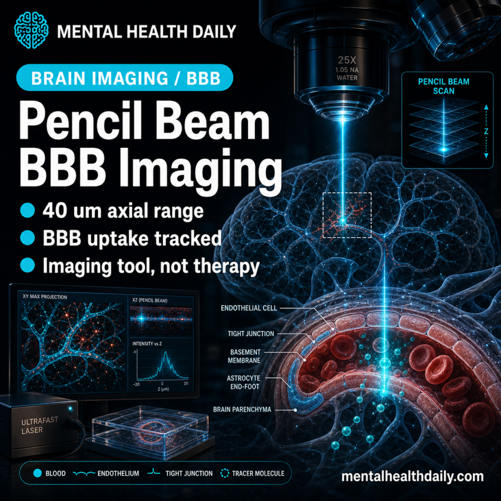
A self-localized ultrafast pencil beam is not a brain treatment, but it could become useful imaging infrastructure for neuroscience and drug-delivery research. In an arXiv preprint, Cao et al. generated a stable ultrafast beam inside a standard multimode fiber and used it for volumetric multiphoton imaging of intact mouse enteric nervous tissue and a live human blood-brain barrier model.1
Research Highlights
- 50-μm fiber self-organization was the optics trick: near critical power, an on-axis overfilled Gaussian beam in a standard multimode fiber reorganized into a stable localized pencil-like beam instead of degrading into random speckle.1
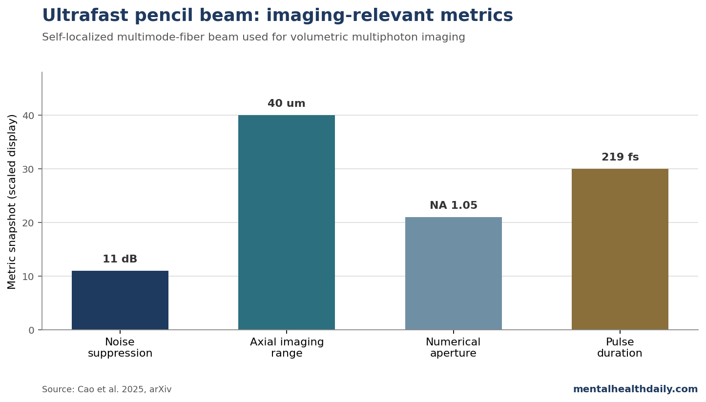
- The reported beam was unusually stable. The localized state achieved 11 dB noise suppression and a sidelobe-suppressed Bessel-like profile.1
- The microscopy payoff was volumetric imaging. The beam supported a continuous 40-μm axial imaging range in a high-NA 1.05 system without customized aperture clearance or adaptive optics.1
- 2 nervous-system demonstrations mattered: the researchers imaged intact mouse enteric nervous systems and tracked transferrin uptake in a live human blood-brain barrier microfluidic model.1
- 3D model tracking is the clinical boundary: this can help observe transport and metabolism in preclinical systems, but it does not establish a diagnostic or therapeutic intervention.
The important distinction: this is an imaging-method paper, not a disease-outcome paper. Its value is that better volumetric microscopy can make neurovascular and cell-delivery biology easier to observe in living systems.
Self-Focusing Became Useful Instead of Destructive
In multimode fibers, intense ultrafast pulses usually create a difficult problem. Nonlinearity, disorder, and mode coupling can scramble the spatial and temporal profile of the beam. Self-focusing near critical power is often treated as a damage or instability risk.1
Cao et al. report a more useful regime. When the input beam was aligned on-axis and well centered, the nonlinear multimode system self-organized into a stable, localized output. The reported setup used 1030-nm pulses, 219-fs temporal duration, and a 50-μm-core silica step-index multimode fiber. Under high peak power, the output shifted from speckled disorder toward a central pencil-like beam.1
A compact method snapshot:
- Input: overfilled on-axis Gaussian beam launched into a standard multimode fiber.
- Pulse parameters: 1030-nm center wavelength and 219-fs duration in the main setup.
- Fiber: 50-μm-core silica step-index multimode fiber.
- Reported optical outcome: self-localized ultrafast pencil beam with 11 dB noise suppression.
- Microscopy outcome: volumetric two-photon imaging with improved sidelobe suppression and resilience to tissue-induced aberration.1
Pencil Beams Extend Focus Without Heavy Sidelobe Noise
The central microscopy problem is speed versus resolution versus tissue scattering. A diffraction-limited Gaussian beam gives tight focus but only at 1 plane at a time. Bessel beams can extend focus axially, which helps volumetric imaging, but sidelobes can excite unwanted out-of-focus fluorescence and reduce contrast.
The self-localized pencil beam aimed to keep the useful extended-focus feature while suppressing sidelobes. The researchers reported that it outperformed conventional Bessel beams in intact mouse enteric nervous tissue, with reduced sidelobes and better resilience to tissue-induced aberration. In a high numerical-aperture system (NA 1.05), it produced efficient signal generation across a continuous 40-μm axial range.1
That combination matters for neuroscience because living tissues are not clean optical phantoms. Brain, gut, vascular, and organoid preparations scatter light, distort wavefronts, move, metabolize, and change during imaging. A beam that preserves volumetric signal under imperfect conditions can be more useful than a beam that looks elegant only in a clean bench setup.
The practical imaging tradeoff is also why the paper spends time benchmarking against Gaussian and Bessel-like approaches. Gaussian scanning can be precise but slow through depth. Bessel beams can cover depth but often pay a price in sidelobes.
A self-localized pencil beam is interesting only if it occupies a better middle ground: enough axial coverage to scan volumes quickly, low enough sidelobes to preserve contrast, and enough robustness to survive real tissue aberration.
That is the reason the enteric nervous system and BBB demonstrations are more persuasive than a purely optical phantom would be. They ask whether the beam remains useful when fluorescent structures are embedded in thick, uneven biological material.
Reader calibration: the beam advance is mainly about optical access, not biological interpretation by itself. A clearer 3D movie can still be misread if the model is immature, the fluorescent marker is nonspecific, or the illumination changes cell behavior. The method improves one bottleneck in live microscopy; it does not remove the need for careful controls around viability, fluorophore behavior, and model validity.
Blood-Brain Barrier Uptake Was the Neurovascular Demonstration
The most MHD-relevant demonstration was the live human blood-brain barrier microfluidic model. The blood-brain barrier is formed by endothelial cells, pericytes, astrocytes, and basement-membrane structures that regulate what moves from blood into brain tissue. It is central to neuropharmacology because many drugs fail not because they cannot bind a target, but because they cannot reach it at useful concentrations.4
The researchers monitored transferrin uptake dynamics in a live human BBB model while combining NAD(P)H/FAD-based metabolic phenotyping with minute-resolved 3D scans. NAD(P)H and FAD are metabolic cofactors whose fluorescence can report cellular redox and metabolic state. Transferrin is relevant because transferrin-receptor pathways are often explored for brain drug-delivery strategies.1,5
The key claim is spatial and temporal heterogeneity. The method revealed different transferrin uptake dynamics across endothelial cells, pericytes, and astrocytes, and variation within cell populations. That is exactly the kind of detail a bulk assay would flatten.
For neuropharmacology, “crosses the BBB” is not a single yes/no property. Transport can differ by receptor avidity, cell type, metabolic state, endothelial maturity, pericyte support, astrocyte signaling, inflammation, and disease model. A method that watches uptake in 3D over minutes can expose failures that an endpoint fluorescence measurement might miss.
The same point applies to metabolic phenotyping. NAD(P)H/FAD signals do not directly diagnose a disease, but they can show whether cells are shifting their redox and energy state while transport is happening. In BBB models, transport and metabolism are intertwined: a stressed or immature barrier may move cargo differently from a healthier one.
That makes the technique most useful for comparing transport conditions inside the same experimental system. For example, researchers could ask whether a receptor-targeted carrier enters endothelial cells but stalls before transcytosis, whether pericyte-supported barriers behave differently from endothelial-only barriers, or whether metabolic stress changes uptake kinetics before barrier leak becomes obvious.
Enteric Nervous System Imaging Was a Useful Stress Test
The mouse enteric nervous system demonstration also matters. The enteric nervous system is a dense neural network embedded in gut tissue, which creates a real biological challenge for volumetric two-photon imaging. The researchers used tdTomato-labeled enteric nervous system tissue and compared the pencil beam with Gaussian and Bessel approaches.1
Enteric nervous system imaging is relevant beyond gut biology. Enteric neurons, autonomic signaling, immune activity, gut-brain communication, and metabolic physiology increasingly overlap with neuropsychiatric and neurodegenerative research. Better volumetric imaging in intact nervous-system tissue can support those fields, even if the method itself is optical engineering.
What the Pencil Beam Does Not Prove
It does not prove clinical diagnostic utility. Imaging a microfluidic BBB model is not the same as imaging the human brain or diagnosing disease in patients.
It does not solve drug delivery. Tracking transferrin uptake helps study delivery pathways, but it does not guarantee that any therapeutic payload will cross the barrier safely or effectively.
It does not remove the need for validation. Preprints need independent replication, comparison across microscopes, tissue types, fluorophores, depths, and laboratories.
It does not make Bessel beams obsolete. The relevant question is which beam architecture works best for a given tissue, depth, speed, phototoxicity tolerance, and resolution target.
Phototoxicity still matters. Faster volumetric imaging is useful only if the illumination does not perturb the biology being measured. Any future biological application needs careful dose, heating, bleaching, and viability checks.
Preclinical Imaging Tool, Not a Blood-Brain Barrier Treatment
For neuroscience: the method could help image 3D biological dynamics in living models where speed, axial range, and contrast all matter.
For blood-brain barrier research: the interesting application is cell-specific transport and metabolism, especially when uptake dynamics differ across endothelial cells, pericytes, and astrocytes.
For drug-delivery work: the tool may improve observation of transport pathways, but delivery efficacy and safety remain separate experimental questions.
For readers: this is not a brain scan for patients. It is a lab microscopy advance that could make preclinical neurovascular models more informative.
Questions About Ultrafast Pencil-Beam Imaging
What is a self-localized ultrafast pencil beam?
It is a stable, narrow, extended optical state generated when an ultrafast pulse self-organizes inside a multimode fiber under carefully aligned high-power conditions.1
Why does sidelobe suppression matter?
Sidelobes can excite unwanted fluorescence outside the intended focal region. Reducing them can improve contrast in volumetric imaging.
What did the blood-brain barrier experiment show?
The researchers tracked transferrin uptake and metabolic signals in a live human BBB microfluidic model, revealing cell-to-cell and within-cell heterogeneity.1
Is this ready for clinical brain imaging?
No. It is a preclinical microscopy method demonstrated in tissue and microfluidic models, not a patient-ready imaging modality.
References
- Self-Localized Ultrafast Pencil Beam for Volumetric Multiphoton Imaging. Cao H, Yu LY, Liu K, et al. arXiv. 2025. doi:10.48550/arXiv.2504.11618
- Video-Rate Volumetric Functional Imaging of the Brain at Synaptic Resolution. Lu R, et al. Nature Neuroscience. 2017;20:620–628. PubMed
- Rapid Mesoscale Volumetric Imaging of Neural Activity with Synaptic Resolution. Lu R, et al. Nature Methods. 2020;17:291–294. PubMed
- Pericytes Regulate the Blood-Brain Barrier. Armulik A, et al. Nature. 2010;468:557–561. doi:10.1038/nature09522
- Transcytosis and Brain Uptake of Transferrin-Containing Nanoparticles by Tuning Avidity to Transferrin Receptor. Wiley DT, Webster P, Gale A, Davis ME. Proceedings of the National Academy of Sciences. 2013;110:8662–8667. doi:10.1073/pnas.1307152110