
People with mild cognitive impairment or Alzheimer’s disease had weaker smell, and the smell tissue inside their noses showed signs of strained antioxidant defenses, a 2026 study of 144 Finnish adults found.1
Research Highlights
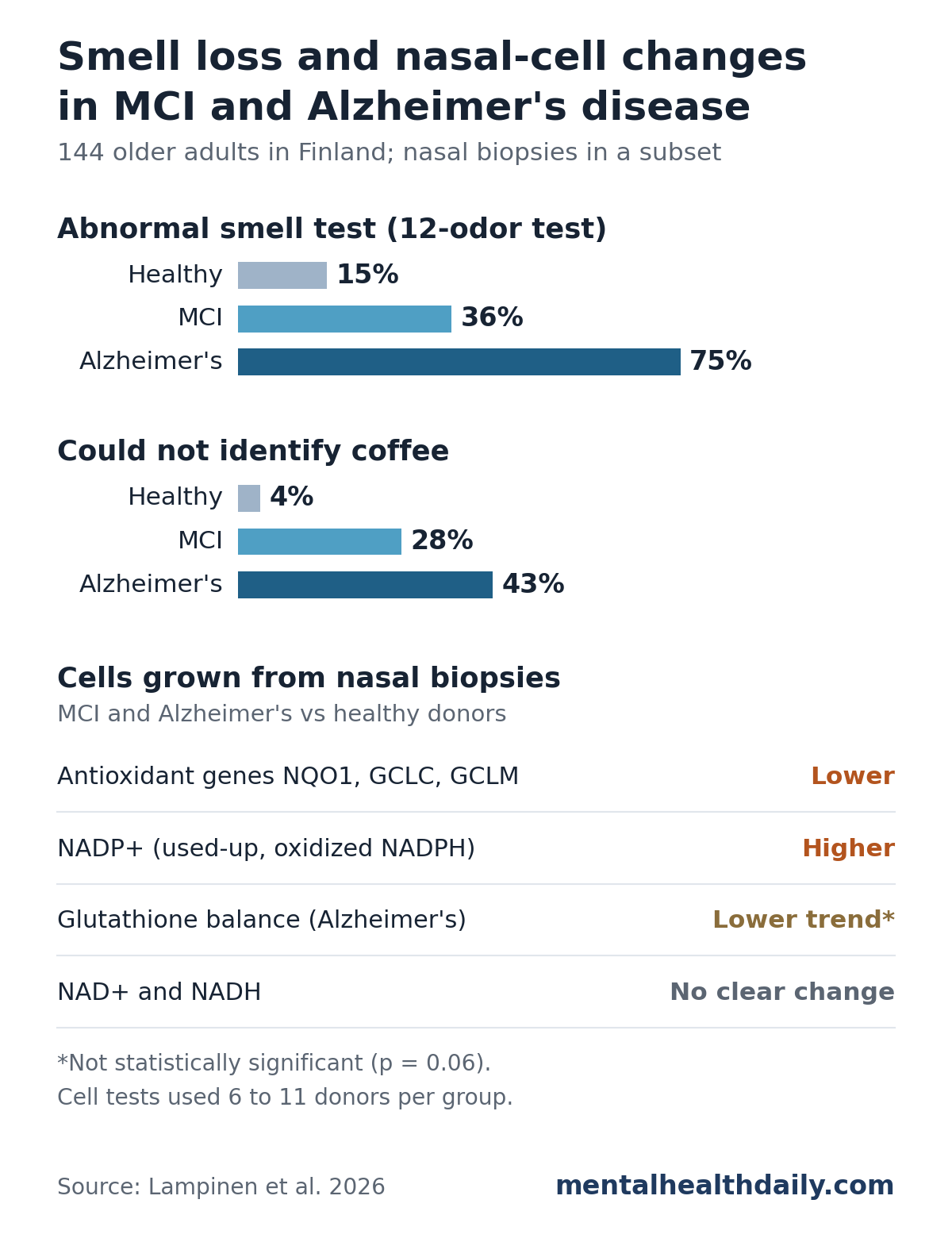
- Smell problems climbed with cognitive decline: 75% of people with Alzheimer’s and 36% with mild cognitive impairment (MCI) had an abnormal 12-odor smell test, vs 15% of cognitively healthy adults.1
- Coffee was the standout odor: 43% of the Alzheimer’s group and 28% of the MCI group could not identify it, vs 4% of healthy adults.1
- Nasal proteins tracked smell scores: of 40 proteins altered in both MCI and Alzheimer’s biopsies, 12 were mitochondrial and 6 were associated with each donor’s smell score.1
- Antioxidant genes were turned down: cells grown from MCI and Alzheimer’s biopsies had lower activity of 3 NRF2-controlled antioxidant genes and more NADP+, the used-up form of a key redox carrier.1
- Small, exploratory molecular work: the protein and redox tests used only 6 to 11 donors per group, and the results are hypothesis-generating.1
Why Researchers Looked Inside the Nose
Trouble identifying smells is an early sign of Alzheimer’s disease. In a classic 2000 study of 90 people with MCI, low smell-test scores (especially when patients were unaware of the problem) predicted who went on to develop Alzheimer’s.2
The olfactory mucosa is the patch of tissue at the roof of the nasal cavity that detects odors. Its nerve cells send fibers straight to the brain’s smell center, and it can be sampled from a living person with a small biopsy, unlike brain tissue.
Earlier MHD coverage looked at smell loss from the brain side, including early nerve-fiber loss in the smell bulb of Alzheimer’s-model mice and MRI patterns linking smell and pTau217 to cognition. This study looked at the nose tissue itself.
What Oxidative Stress and Redox Balance Mean
Cells constantly run chemical reactions that pass electrons from one molecule to another. Redox balance (short for reduction-oxidation) is the cell’s running tally of that electron traffic.
A side effect of making energy in mitochondria is a stream of reactive oxygen molecules, often called free radicals. When they outpace the cell’s cleanup systems, they damage proteins, fats and DNA. That imbalance is oxidative stress.
The study focused on a few of the cell’s defenders:
- NRF2: a master switch protein that turns on dozens of antioxidant and cleanup genes when stress rises.
- NQO1, GCLC and GCLM: three of those NRF2-controlled genes. GCLC and GCLM build glutathione; NQO1 neutralizes reactive compounds.
- Glutathione: the cell’s main antioxidant molecule. Its reduced form (GSH) is ready to work; its oxidized form (GSSG) has been used.
- NADPH and NADP+: electron carriers. NADPH supplies the power to recharge glutathione; NADP+ is the spent form left behind.
NRF2 problems have been seen in the brain as well. In Alzheimer’s hippocampus tissue, NRF2 tends to stay outside the cell nucleus, where it cannot switch genes on, even though oxidative damage is present.3
Who Was in the Finnish Smell and Biopsy Study
This was a human-only study, run at Kuopio University Hospital and the University of Eastern Finland. No animals were used; the cells in the lab tests were grown from the participants’ own nasal biopsies.1
- Smell testing: 144 adults (53 cognitively healthy, 47 with MCI, 44 with mild Alzheimer’s dementia), average age about 70, took the 12-odor Sniffin’ Sticks identification test.
- Biopsies: 16 people per group gave a small sample of olfactory mucosa from the upper nasal septum.
- Protein analysis: 6 biopsies per group were analyzed for thousands of proteins at once (shotgun proteomics).
- Cell tests: cells grown from biopsies were checked for NRF2 target genes (10 to 11 donors per group), redox molecules (6 per group) and microRNAs.
MCI here meant measurable memory problems without loss of everyday independence. In the protein-analysis donors, a blood marker of Alzheimer’s tau pathology (plasma pTau217) was highest in the Alzheimer’s group and intermediate in MCI, which supported the group labels.1
Coffee and Fish Were Hardest to Identify in MCI and Alzheimer’s
Overall, 75% of the Alzheimer’s group had reduced or absent smell (hyposmia or anosmia), compared with 36% of the MCI group and 15% of healthy adults.1
Ten of the 12 odors separated the Alzheimer’s group from healthy adults. Cinnamon and clove did not separate any groups. Four odors were missed significantly more often in MCI too:
- Coffee: missed by 4% of healthy adults, 28% with MCI and 43% with Alzheimer’s.
- Fish: 6%, 30% and 41%.
- Banana: 9%, 28% and 48%.
- Lemon: 40%, 64% and 73%.
Orange was missed by 45% of the Alzheimer’s group vs 11% of healthy adults, though the MCI difference (21%) was not significant. A machine-learning analysis picked coffee, orange, banana, fish and lemon as the most useful set for telling the 3 groups apart.1
Lemon is a weak marker on its own: 40% of healthy adults also missed it.
Nasal Tissue Proteins in MCI and Alzheimer’s Pointed to Mitochondria
Across the 18 analyzed biopsies, researchers identified 6,031 proteins. Compared with healthy tissue, 184 differed in MCI and 901 differed in Alzheimer’s; 40 changed in both.1
Mitochondria stood out. Of those changed proteins, 116 of 901 in Alzheimer’s, 48 of 184 in MCI and 12 of the 40 shared ones belong to mitochondria, the cell’s energy plants and main source of free radicals. In MCI, the altered pathways centered on cellular respiration.1
Six shared proteins tracked smell scores. VAMP5, COQ7, CTSB, RPS21, CLTA and HSD17B10 were associated with how well each donor did on the smell test. COQ7 helps make coenzyme Q10 in mitochondria, and HSD17B10 is a mitochondrial enzyme that binds amyloid-beta.1
NRF2 markers shifted. Fifty-six altered proteins in Alzheimer’s tissue and 16 in MCI tissue are known markers of NRF2 activity. Several proteins that NRF2 directly controls were lower in Alzheimer’s tissue, and one of them, CD99, was also lower in MCI.1
Cells From MCI and Alzheimer’s Biopsies Showed Weaker Antioxidant Signaling
To test redox balance directly, the team grew cells from the biopsies in the lab. Several findings lined up with the tissue results:1
- Lower antioxidant gene activity. NQO1, GCLC and GCLM were significantly lower in cells from both MCI and Alzheimer’s donors than in cells from healthy donors.
- More spent NADP+. NADP+ was significantly higher in MCI and Alzheimer’s cells, and highest in MCI. The researchers read this as cells using up NADPH faster or needing more of it.
- A glutathione trend only. Total glutathione did not differ. The ratio of ready-to-use to used glutathione was slightly lower in Alzheimer’s cells, but the difference was not significant (p = 0.0617).
- No change in NAD+ or NADH, a separate electron-carrier pair used mainly in energy production.
A microRNA that targets NRF2 was higher. MicroRNAs are short RNA pieces that turn down specific genes. One called miR-142-5p, which is predicted to target the NRF2 gene, was raised in Alzheimer’s cells (log2 fold change 1.78), and 67 of the altered biopsy proteins are listed as its targets in interaction databases.1
Together, the results suggest NRF2 defenses are dampened in the smell tissue of people with cognitive decline, at a stage when smell is already slipping.
Limitations of This Nasal Biopsy Study
Evidence strength: a cross-sectional, single-center human study with small molecular subsets. It shows associations; it cannot show that redox changes cause smell loss or Alzheimer’s.
- Tiny molecular groups. Protein and redox tests used 6 donors per group; gene tests used 10 to 11.
- No multiple-testing correction. Protein and microRNA hits were called on nominal p-values. With thousands of proteins tested, some will be chance findings, and the researchers call these results exploratory.
- Oxidative damage itself was not measured. Redox status was inferred from genes and carrier molecules, not from damaged DNA or fats.
- Lab-grown cells. Cultured cells may not behave exactly like tissue in a living nose, and biopsies mix smell tissue with ordinary nasal lining.
- Uneven groups. 70% of healthy controls were women vs 36% in MCI and 43% in Alzheimer’s, though the protein analysis adjusted for age and sex.
- Nose is not brain. The nose is exposed to air pollutants and infections, which can also raise oxidative stress there.
What This Means for Smell Tests and Alzheimer’s Risk
A smell test alone cannot diagnose Alzheimer’s. Many healthy older adults also miss odors, and other conditions affect smell. The researchers suggest odor-specific results could someday be combined with blood biomarkers to flag people at higher risk.1
The redox findings point to a biological process worth tracking, not a treatment. No antioxidant, supplement or NRF2-targeting drug was tested here. Larger studies with more biopsies will need to confirm which of these nasal changes hold up.
Anyone noticing a new, persistent loss of smell along with memory changes should raise it with a doctor, since both have many possible causes.
References
- Lampinen R, Afonin AM, Mussalo L, et al. Cellular redox status is linked to olfactory alterations in cognitive decline. Redox Biology. 2026;97:104393. doi:10.1016/j.redox.2026.104393
- Devanand DP, Michaels-Marston KS, Liu X, et al. Olfactory deficits in patients with mild cognitive impairment predict Alzheimer’s disease at follow-up. American Journal of Psychiatry. 2000;157(9):1399–1405. doi:10.1176/appi.ajp.157.9.1399
- Ramsey CP, Glass CA, Montgomery MB, et al. Expression of Nrf2 in neurodegenerative diseases. Journal of Neuropathology & Experimental Neurology. 2007;66(1):75–85. doi:10.1097/nen.0b013e31802d6da9