

In 53 South Korean women with memory decline, higher Alzheimer’s blood markers were linked to a thinner brain cortex only in women with at least 7 years of schooling. In women with 6 or fewer years, cortical thickness did not track the markers.1
Research Highlights
- Thinning showed up only with more schooling: among women with 7 to 16 years of education, a higher blood p-tau/amyloid ratio went with a thinner cortex in all 5 brain regions measured (p = 0.005 to 0.028).1
- No link in women with 0 to 6 years: their cortical thickness did not track the blood markers in any region.1
- Preserved thickness did not mean better thinking scores: cortical thickness was not linked to Mini-Mental State Examination (MMSE) scores in these models.1
- Women vs. men: across all 80 patients, women had a higher p-tau/amyloid ratio yet a thicker frontal cortex than men.1
- Small, single-clinic snapshot: a cross-sectional study of 80 patients (58 women, 22 men) published in the Journal of Clinical Neurology.1
How Researchers Measured Alzheimer’s Markers and Brain Thickness
Yeonsil Moon et al. enrolled 80 patients who came to a Seoul memory clinic between November 2021 and March 2022 with mild cognitive impairment or dementia. Their average age was 77.9, and they had about 7.4 years of schooling on average.1
Each patient had a blood draw, a brain MRI, and standard cognitive screening. Three measurements carried the study:
- Blood biomarkers: amyloid-beta 42 (a protein fragment that clumps into Alzheimer’s plaques; blood levels tend to fall as plaques build in the brain) and p-tau181 (a modified form of tau, the protein that forms tangles inside nerve cells). The main marker was the p-tau/amyloid ratio, which rises as Alzheimer’s pathology increases.
- Cortical thickness: the cortex is the brain’s folded outer layer of nerve cells. As Alzheimer’s kills neurons, the cortex thins, so MRI thickness is a common way to gauge brain damage. The team measured frontal, parietal, temporal, occipital, and insular regions.
- Cognition: the MMSE, a 30-point bedside screening test, and the Clinical Dementia Rating–Sum of Boxes, a measure of how much memory problems affect daily life.
Education was split at 6 years. Many women now in their late 70s in South Korea had little chance to study past elementary school, so 0 to 6 years marks finishing elementary school, and 7 to 16 years marks middle school or beyond.1
Why Education Is Used to Measure Cognitive Reserve
Cognitive reserve is the brain’s ability to keep working well despite damage. People with more reserve can carry more Alzheimer’s pathology before symptoms show, and they are often diagnosed later.2
Reserve cannot be measured directly, so researchers use stand-ins such as years of schooling. In this cohort, schooling was also heavily linked to sex: 93% of the low-education group were women, and 86% of the men had 7 or more years of education.1
Because low education and female sex overlapped so heavily, the researchers ran their main test only in women. That analysis included the 53 women with complete MRI and blood data, adjusted for age.1
More-Educated Women Showed Thinning as Alzheimer’s Markers Rose
The link between blood markers and cortical thickness differed by education in every region tested. The statistical interaction was strongest in the occipital and temporal cortex (both p = 0.026) and weakest, though still significant, in the parietal cortex (p = 0.049).1
Splitting the women by schooling showed what drove that interaction:
- 7 to 16 years of school: a higher p-tau/amyloid ratio went with a thinner cortex in all 5 regions, the pattern doctors expect as Alzheimer’s advances.
- 0 to 6 years of school: no significant link in any region. The estimates even pointed slightly toward a thicker cortex, but none reached significance (p = 0.152 to 0.311).
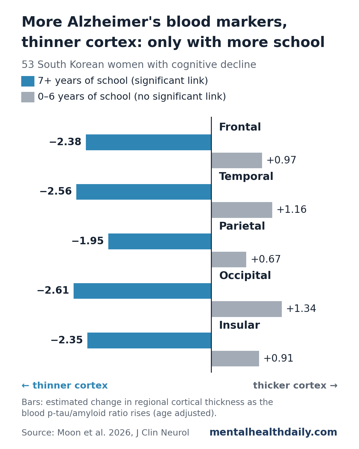
Results held when the team tried other education cutoffs (0 to 3, 4 to 9, and 10 or more years) and when they treated schooling as a continuous number of years.1
A Thicker Cortex Did Not Mean Better Thinking Scores
If preserved thickness were true protection, it should have shown up as better cognitive scores. It did not: in the women-only models, cortical thickness was not linked to MMSE scores in any region, so the education difference in thickness did not carry through to cognition.1
The researchers read this as a delayed structural response rather than real resilience. The cortex in less-educated women may simply not show the damage yet. They suggest 2 possible reasons, neither tested here:
- Inflammation and swelling. Early in Alzheimer’s, reactive support cells (astrocytes) and inflammation could swell brain tissue and mask early neuron loss on MRI.
- Blood vessel damage. Vascular problems, more common with lower socioeconomic status, could cause subtle swelling that keeps cortical volume up despite ongoing damage.
Clinical implication: in women with little formal education, a normal-looking MRI could underestimate Alzheimer’s damage and delay diagnosis. The researchers recommend reading structural MRI results with education in mind; larger studies still need to confirm the pattern.1
Women Had Higher Alzheimer’s Markers but a Thicker Frontal Cortex Than Men
Across all 80 patients, the log-transformed p-tau/amyloid ratio was higher in women than in men (−0.37 vs. −0.53, p = 0.026). Women also had a thicker frontal cortex (4.2 vs. 3.2 mm, p = 0.049), with similar but nonsignificant trends in the other 4 regions.1
Raw MMSE scores were lower in women (16.8 vs. 21.2), but scores adjusted for age and education did not differ by sex. Formal tests found no sex-by-education interaction for the blood ratio or MMSE.1
A thicker cortex in women despite equal or greater pathology fits earlier imaging work. In a 2022 analysis of the US-based Alzheimer’s Disease Neuroimaging Initiative, women had greater cortical thickness in many regions, held thickness steadier from normal cognition to mild cognitive impairment, and then showed sharper declines on the way to Alzheimer’s dementia.3
Limitations of This Education and Brain-Thickness Study
This is a cross-sectional study: it captured one moment in time and can show links, not cause and effect or what happens to these women later. Other limits bear directly on the findings:
- Small sample: 80 patients overall and 53 in the main analysis, from a single memory clinic.
- Sex and education were tangled: with only 22 men, nearly all well educated, the study could not test whether the same education pattern applies to men. The sex differences are descriptive only.
- Education stands in for more than school: in this generation, schooling also reflects childhood family income and other life advantages that shape the brain.
- Only screening tests for cognition: the MMSE may be too blunt to catch subtle benefits in memory or planning that a thicker cortex could support.
- Mechanisms are untested: the swelling and vascular explanations are the researchers’ hypotheses.
Long-term follow-up in larger, better-balanced groups is needed to see whether the preserved cortex in less-educated women later thins quickly once a threshold is crossed.1
References
- Moon Y, Jeon HJ, Kim HJ, et al. Education moderates the biomarker–cortical-thickness relationship in females with cognitive decline: a descriptive study with sex comparisons. Journal of Clinical Neurology. 2026;22(5):552–559. doi:10.3988/jcn.2025.0686
- Stern Y. Cognitive reserve in ageing and Alzheimer’s disease. Lancet Neurology. 2012;11(11):1006–1012. doi:10.1016/S1474-4422(12)70191-6
- Cieri F, Zhuang X, Cordes D, et al. Relationship of sex differences in cortical thickness and memory among cognitively healthy subjects and individuals with mild cognitive impairment and Alzheimer disease. Alzheimer’s Research & Therapy. 2022;14:36. doi:10.1186/s13195-022-00973-1