
Blocking immune cells from entering the brain with the multiple sclerosis drug natalizumab did not significantly change brain glutamate in people with first-episode psychosis. A PET immune marker also showed no link to glutamate in the same brain region.1
Research Highlights
- No glutamate change after natalizumab: in 32 patients given 3 monthly infusions, anterior cingulate glutamate was 0.227 units higher at follow-up, a nonsignificant difference (p = 0.139). The predicted drop did not appear.1
- No link at baseline: in 100 people with both scans, TSPO binding (a PET marker of brain immune cells) did not track glutamate (β = −0.003, p = 0.553), in patients or in healthy controls.1
- Glutamate did not follow symptoms: changes in glutamate were unrelated to changes in symptom scores after natalizumab (p = 0.948).1
- Sample: 108 people with usable brain spectroscopy (68 with first-episode psychosis, 40 controls) at 4 London early-intervention services; Neuropsychopharmacology, 2026.1
- Mixed design: the treatment arm pooled a double-blind randomized phase with an open-label phase added after COVID-19 disrupted the study.1
Why Researchers Expected Brain Immune Cells to Raise Glutamate in Schizophrenia
Two separate lines of research point to problems in schizophrenia. One involves the brain’s immune system; the other involves glutamate signaling.
- Microglia are the brain’s resident immune cells. Post-mortem studies have found more of them, and more of a protein they make when inflamed, in some people with schizophrenia.1
- Glutamate is the brain’s main excitatory (“go”) chemical messenger. Higher glutamate in the anterior cingulate cortex (ACC), a midline frontal region involved in attention, motivation and emotion, has been linked to worse symptoms and a poorer response to antipsychotics.1
Animal and cell studies connect the two. Activated microglia can push extra glutamate out of cells through a cystine-glutamate exchanger, and can release TNF-α, an inflammatory signal that stops neighboring astrocytes from clearing glutamate.1
If that happens in psychosis, overactive microglia could be raising glutamate, and calming the immune system might bring glutamate down. Hindley et al. tested both steps in living patients.
How the Natalizumab PET/MRS Study Measured Immune Activity and Glutamate
Each participant lay in a combined PET-MRI scanner for about an hour, so both measures were taken in one session.1
- PET and TSPO: Positron emission tomography (PET) uses a small dose of radioactive tracer to map where a target protein sits. This study’s tracer, [18F]DPA-714, binds translocator protein (TSPO), which microglia produce in large amounts when inflamed. More binding is read as more immune activity.
- MRS and glutamate: Proton magnetic resonance spectroscopy (MRS) uses an MRI scanner to read the chemical makeup of a small block of tissue. Here it was a 2 × 2 × 2 cm cube in the ACC. It measures glutamate and Glx (glutamate plus glutamine, a closely related molecule that is hard to separate at this field strength).
- Natalizumab: a monoclonal antibody (a lab-made immune protein) approved for multiple sclerosis. It blocks immune cells from crossing blood vessel walls into the brain, and it lowered TSPO PET signal in multiple sclerosis. Patients received 300 mg by IV infusion once a month for 3 months.
Who took part: adults aged 18 to 50 with first-episode psychosis, ill for under 5 years and still symptomatic on a stable antipsychotic (not clozapine), plus healthy controls. About 65% of patients had a schizophrenia diagnosis. Average total score on the Positive and Negative Syndrome Scale (PANSS), a clinician-rated measure of psychosis symptoms, was 57.3, which corresponds to “mildly ill.”1
Treatment arms: 48 patients were randomized 1:1 to natalizumab or saline placebo, double-blind. After the pandemic disrupted recruitment, 18 more patients received open-label natalizumab. After scan dropouts and quality checks, 32 natalizumab recipients (17 randomized, 15 open-label) and 18 placebo recipients had usable before-and-after spectroscopy.1
No Link Between the TSPO Immune Marker and ACC Glutamate at Baseline
Of 74 patients and 42 controls recruited, 68 and 40 had usable MRS, and 61 and 39 also had usable PET.1 In those 100 people, TSPO binding in the ACC did not predict ACC glutamate or Glx:
- Whole sample: glutamate β = −0.003 (p = 0.553); Glx β = −0.004 (p = 0.263)
- Patients only (61): glutamate β = −0.009 (p = 0.244)
- Controls only (39): glutamate β = 0.001 (p = 0.865)
These slopes are essentially flat. Adjusting for TSPO genotype, age, sex and smoking did not change them, and TSPO in other regions (total, frontal and temporal gray matter) showed no link with ACC glutamate either.1
Group differences: patients had slightly lower ACC Glx than controls (11.1 vs 11.6 units, p = 0.040). ACC glutamate (p = 0.151) and ACC TSPO binding (p = 0.199) did not differ between groups.1 In the same cohort, TSPO binding was higher in patients across total and temporal gray matter, but not in the frontal lobe.2
Natalizumab Did Not Significantly Change Anterior Cingulate Glutamate
The treatment test was simple: did glutamate fall in patients who received natalizumab? It did not.1
- Glutamate: 0.227 units higher at follow-up (p = 0.139)
- Glx: 0.296 units higher (p = 0.083)
- Only patients who got all 3 infusions: glutamate 0.281 higher (p = 0.095); Glx 0.391 higher (p = 0.062)
- Placebo, for comparison: glutamate −0.046 (p = 0.781); Glx +0.257 (p = 0.303)
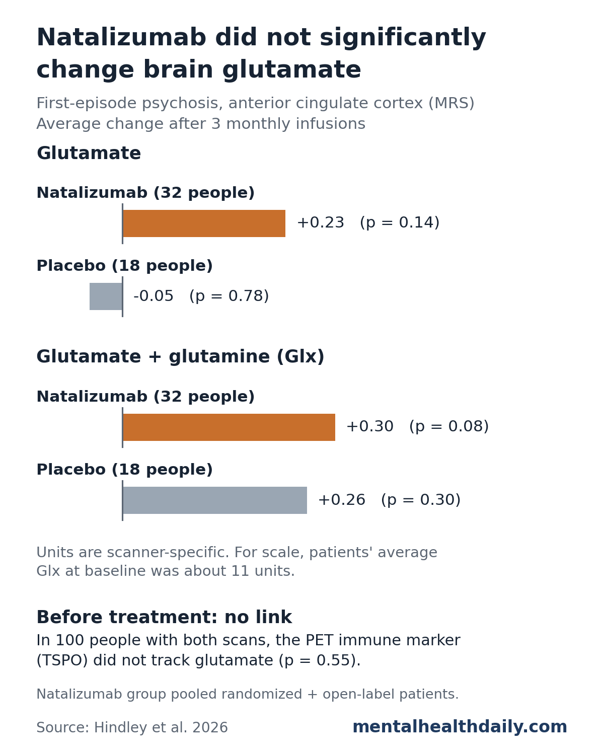
None of these changes reached significance. In the natalizumab group the small shifts pointed upward, the opposite of the predicted drop, and they were small next to baseline Glx levels of about 11 units.1
Because natalizumab did not lower glutamate, the researchers did not go on to their planned between-group comparison with placebo. Adjusting for age, sex and smoking left the result unchanged.1
Symptoms and glutamate: in the parent report from this trial, PANSS total scores improved modestly within the natalizumab group (mean change −3.7 points, Cohen’s d = 0.40).2 In the 2026 analysis, those symptom changes were unrelated to glutamate changes (PANSS total β = −0.690, p = 0.948).1
Natalizumab Did Not Lower the TSPO Signal Either
The parent report found no significant change in TSPO binding after natalizumab in total, frontal or temporal gray matter.2 That weakens the treatment test as a check on the microglia idea.
If the drug never calmed the immune activity the scans measure, glutamate had no reason to fall. The researchers give 2 possible explanations:1
- The drug did not lower the immune marker. Natalizumab did not reduce TSPO in these patients, so the glutamate question was never fully put to the test.
- There is no link to find. The flat baseline result suggests TSPO binding and ACC glutamate are simply unrelated in early psychosis.
Earlier TSPO and Glutamate Studies in Psychosis
The only earlier direct test agrees. A Toronto study compared hippocampal glutamate (by MRS) with TSPO PET in 25 people at clinical high risk for psychosis, 16 with first-episode psychosis and 31 healthy volunteers. It found no associations between glutamate metabolites and glial activation.3
TSPO findings in schizophrenia depend on how they are measured. A meta-analysis of 12 studies (190 patients, 200 controls) found higher TSPO binding in schizophrenia with one quantification method (binding potential; Hedges’ g = 0.31, p = 0.03) but no difference with another (volume of distribution; g = −0.22, p = 0.29).4
The ACC may not be the best place to look. A meta- and mega-analysis of MRS studies found the most robust glutamate differences in schizophrenia in the thalamus and basal ganglia, with a small reduction in medial frontal glutamate (g = −0.15).5 Hindley et al. suggest future studies scan those regions too.1
Limitations of This Natalizumab Imaging Study
The findings come with clear limits:1
- Mixed treatment design: the natalizumab group combined blinded randomized patients and open-label patients, and the main test was a before-and-after comparison within that group.
- Power: the treatment test was sized to detect a change of about Cohen’s d = 0.72, so a smaller real change could be missed. The placebo group was too small to test on its own.
- Data loss: scan quality checks and dropouts removed participants at each stage; 50 of the 66 patients who started infusions made the final longitudinal analysis.
- TSPO is an imperfect microglia marker: astrocytes and blood vessel cells also make it, and it says little about what microglia are actually doing.
- Coarse glutamate measure: 1 ACC voxel averages millions of synapses, so local changes at the synapse could be missed. Units are scanner-specific and cannot be compared across sites.
- A mildly ill, medicated sample: all patients were on antipsychotics, and a more symptomatic or immune-selected group might behave differently.
What the Results Mean for Immune Treatments in Schizophrenia
- Separate problems, for now: in early psychosis, raised TSPO binding and ACC glutamate changes look like distinct processes rather than one chain of cause and effect.
- Immune ideas are not ruled out: the study tested 1 region, 1 immune marker and 1 drug that did not lower that marker. Other immune pathways or brain regions could still matter.
- Not a treatment result: natalizumab is not approved for psychosis or schizophrenia, and this imaging study was not built to show whether it helps symptoms.
- Next steps: the researchers suggest enrolling patients with signs of immune activation or glutamate problems, and using more specific microglia markers.
References
- Hindley G, Mizuno Y, Beck K, et al. Does a monoclonal antibody targeting immune cells affect glutamatergic level in schizophrenia? A multimodal PET/MRS brain imaging study. Neuropsychopharmacology. 2026;51:2083–2092. doi:10.1038/s41386-026-02466-3
- Mizuno Y, Carreira Figueiredo I, Pillinger T, et al. Immune alterations in schizophrenia and the effects of a therapeutic antibody: a neuroimaging study. Brain. 2025. doi:10.1093/brain/awaf455
- Shakory S, Watts JJ, Hafizi S, et al. Hippocampal glutamate metabolites and glial activation in clinical high risk and first episode psychosis. Neuropsychopharmacology. 2018;43:2249–2255. doi:10.1038/s41386-018-0163-0
- Marques TR, Ashok AH, Pillinger T, et al. Neuroinflammation in schizophrenia: meta-analysis of in vivo microglial imaging studies. Psychological Medicine. 2019;49:2186–2196. doi:10.1017/S0033291718003057
- Merritt K, McCutcheon RA, Aleman A, et al. Variability and magnitude of brain glutamate levels in schizophrenia: a meta and mega-analysis. Molecular Psychiatry. 2023;28:2039–2048. doi:10.1038/s41380-023-01991-7