
Long-lasting inflammation in the placenta was linked to differences in how fetal brain regions work together, in a 2026 MRI study of 105 pregnancies.
One of those differences, a weaker link between the cerebellum and the brain’s visual areas, went along with higher parent-reported autism trait scores at age 3.1
Research Highlights
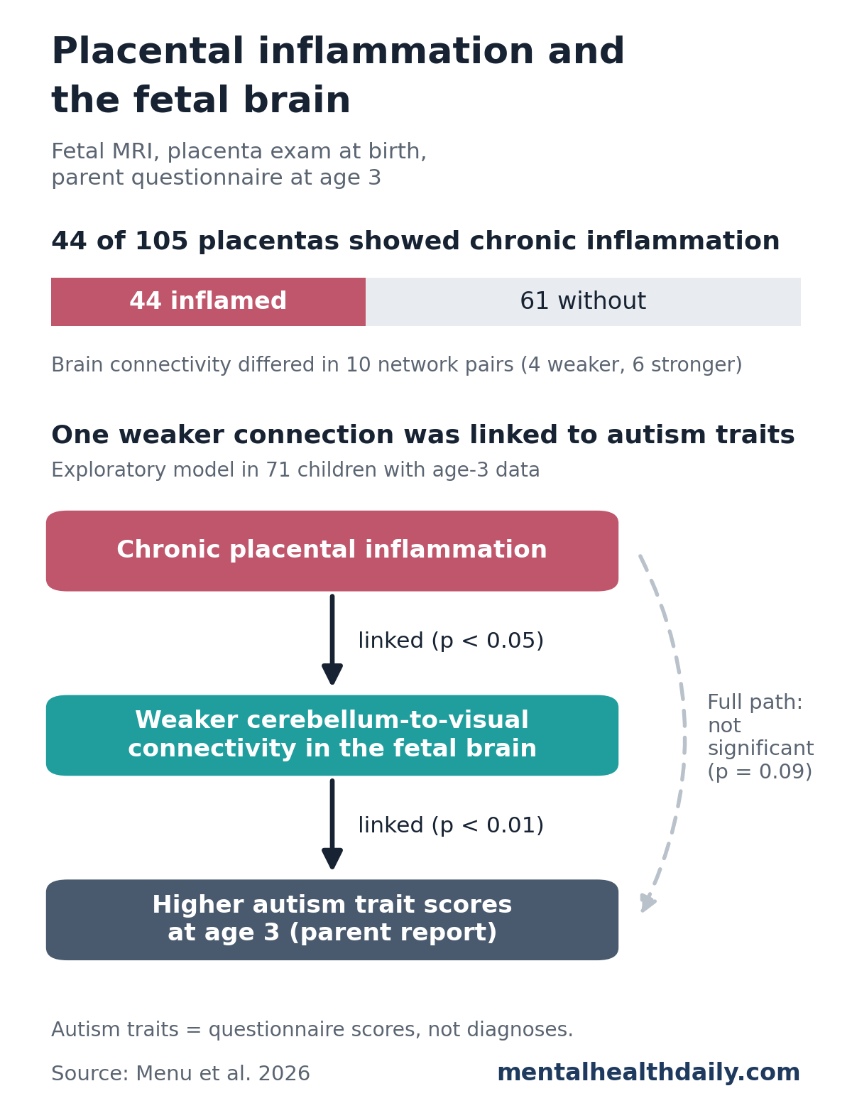
- Common finding: 44 of 105 placentas (41.9%) showed at least one type of chronic inflammation when pathologists examined them after delivery.1
- Different fetal brain wiring: fetuses exposed to placental inflammation had different connectivity in 10 pairs of brain networks — 4 weaker and 6 stronger — compared with unexposed fetuses.1
- Cerebellum–visual link stood out: weaker connectivity between the cerebellum and the medial visual network was the most consistent prenatal predictor of autism trait scores at age 3.1
- Full pathway not confirmed: the step from inflammation through brain connectivity to autism traits did not reach statistical significance (p = 0.09), and inflammation had no direct link to autism traits.1
- Traits, not diagnoses: autism traits came from a 13-item parent questionnaire, and no child in the study was reported as diagnosed with autism.1
What Is Chronic Placental Inflammation?
The placenta is the organ that passes oxygen and nutrients from mother to fetus. It also sits where the mother’s immune system meets tissue that is partly foreign to it, because half of the fetus’s genes come from the father.
Chronic placental inflammation (CPI) means maternal immune cells, such as T cells and plasma cells, have moved into placental tissue, an inflammatory process that builds over the course of pregnancy. It can follow an infection, or it can reflect the mother’s immune system reacting against fetal tissue, a process pathologists call maternal anti-fetal rejection.2
Pathologists recognize 3 main types, depending on which part of the placenta is affected:2
- Villitis of unknown etiology (VUE): inflammation in the villi, the tiny branching structures where fetal blood picks up oxygen and nutrients (15.2% of the study sample)
- Chronic chorioamnionitis: inflammation in the membranes that form the amniotic sac (26.7%)
- Chronic deciduitis: inflammation in the decidua, the lining of the uterus where the placenta attaches (20%)
These lesions often occurred together, so the researchers grouped any of the 3 as one exposure. In this study, the inflammation could only be identified after birth, when 2 perinatal pathologists who did not know the brain results examined each placenta under a microscope.1
How Researchers Measured Fetal Brain Development
Iris Menu et al. recruited 105 pregnant women during routine prenatal visits at the Detroit Medical Center. Each fetus had a resting-state functional MRI scan between 24 and 38 weeks of pregnancy (30.7 weeks on average).1
Resting-state MRI tracks slow rises and falls in blood oxygen across the brain while no task is being done. When 2 regions rise and fall in step, they are considered functionally connected, a measure called resting-state functional connectivity. Researchers divided each fetal brain into 197 regions and grouped them into 15 networks.1
Three years later, mothers filled out the Child Behavior Checklist (CBCL), a widely used parent questionnaire. Its 13-item Autism Spectrum Problems scale asks about social give-and-take, communication and repetitive behaviors. It is a screening tool that flags children who may need further evaluation, not a diagnostic test.1
Study snapshot:
- Sample: 105 mother–fetus pairs; mothers aged 18 to 41 (average 25.7), 85.7% Black
- Groups: 44 with placental inflammation, 61 without; the groups were similar in fetal age at scan, birth weight, preterm birth, sex, income and education
- Age-3 follow-up: autism trait scores available for 71 children (67.6%)
Placental Inflammation Was Linked to 10 Fetal Network Differences
Compared with unexposed fetuses, fetuses whose placentas showed chronic inflammation had different connectivity in 10 pairs of brain networks. The differences held up after the researchers also accounted for maternal age and the child’s sex.1
- 4 weaker connections: left prefrontal–right operculum, cerebellum–cingulate, cerebellum–medial visual, and ventromedial prefrontal–right motor
- 6 stronger connections: mostly links among visual, prefrontal, motor and cerebellar networks, plus left temporal–ventromedial prefrontal
The cerebellum, at the back of the brain, coordinates movement and also supports thinking and emotional regulation. It grows quickly around birth, and cerebellar differences are among the most consistent brain imaging findings in autism.1
Menu et al. suggested the weaker links may reflect disrupted pathways, while the stronger ones could be the developing brain rerouting around the inflammation. The study did not test either idea directly.1
Weaker Cerebellum–Visual Connectivity Went With Higher Autism Trait Scores
To see which fetal connections best predicted autism trait scores at age 3, the researchers used a regression model that keeps only the strongest predictors. Across repeated resampling runs, cerebellum–medial visual connectivity was selected 95.6% of the time.1
The model’s overall accuracy was modest. Fetal connectivity explained about 15% of the variation in autism trait scores on average, with a 95% confidence interval from 0% to 43%.1
An exploratory pathway model in the 71 children with age-3 data then tested the full chain:
- Inflammation to connectivity: placental inflammation was linked to weaker cerebellum–visual connectivity (standardized β = −0.25, p < 0.05).
- Connectivity to traits: weaker connectivity was linked to higher autism trait scores (β = −0.32, p < 0.01).
- Full pathway: the indirect effect of inflammation on autism traits through connectivity was not significant (β = 0.08, p = 0.09), and there was no direct link between inflammation and autism traits (p = 0.91).
The researchers called the pathway result hypothesis-generating. Other factors, such as genetic risk or chronic stress, could affect both brain connectivity and autism traits on their own.1
How the Findings Fit Earlier Research on Inflammation and Autism
Infection and inflammation during pregnancy have been linked to autism for years. A 2016 meta-analysis of 15 studies with more than 40,000 autism cases found maternal infection was linked to modestly higher odds of autism in offspring (OR = 1.13), and somewhat higher odds when the infection required a hospital stay (OR = 1.30).3
Most earlier human work relied on maternal blood markers or brain scans taken after birth. This study looked directly at the fetal brain and used placental tissue to measure inflammation, which Menu et al. described as the first human evidence of this kind.1
Limits of This Fetal MRI Study
An observational study like this one can show what goes together, but it cannot show that placental inflammation changes the fetal brain or causes autism traits. Specific limits:
- Questionnaire, not diagnosis: autism traits came from a parent-report screener at age 3; no observational or diagnostic assessment was done.
- Missing follow-up: 34 of 105 children (32%) had no age-3 data.
- Unmeasured factors: the study lacked full data on maternal infection, antibiotic use and autoimmune conditions, which could affect both the placenta and the fetal brain.
- Lumped lesion types: the 3 types of inflammation have different biology but were analyzed as one group.
- Overlapping data: the brain network map was built from 168 fetal scans that included the 105 studied, so the network boundaries were not fully independent of the results.
- One community: participants were mostly Black, lower-income Detroit families facing high stress, so results may differ in other groups.
For expectant parents: chronic placental inflammation was common in this sample, and the study did not show that it leads to autism. In this study it was found only by examining the placenta after birth, and the findings do not point to any test or prevention step during pregnancy.
References
- Menu I, Ji L, Chen B, et al. Associations between chronic placental inflammation, fetal brain development and later autism traits. Brain, Behavior, & Immunity – Health. 2026;57:101351. doi:10.1016/j.bbih.2026.101351
- Kim CJ, Romero R, Chaemsaithong P, Kim JS. Chronic inflammation of the placenta: definition, classification, pathogenesis, and clinical significance. American Journal of Obstetrics and Gynecology. 2015;213(4 Suppl):S53–S69. doi:10.1016/j.ajog.2015.08.041
- Jiang HY, Xu LL, Shao L, et al. Maternal infection during pregnancy and risk of autism spectrum disorders: a systematic review and meta-analysis. Brain, Behavior, and Immunity. 2016;58:165–172. doi:10.1016/j.bbi.2016.06.005