
A 2026 MRI study involving 86 adults found that obsessive-compulsive disorder (OCD) and alcohol use disorder differed from each other in the same frontal, cingulate, and brainstem regions, but in opposite directions: gray matter tended to be larger in OCD and smaller in alcohol use disorder.1
Research Highlights
- Opposite gray matter pattern: 11 of 126 gray-matter regions differed by group after strict correction for multiple tests, and OCD volumes were larger than alcohol use disorder volumes in all 11.1
- Both groups differed from controls: compared with healthy controls, the OCD group had a larger brainstem (p = 0.0001), while the alcohol use disorder group had smaller frontal and cingulate regions, such as the right frontal operculum (p = 0.0014).1
- No clear white matter differences: none of the 68 white-matter regions differed between groups.1
- No link to symptom severity: regional volumes did not track OCD severity or alcohol craving scores.1
- Small snapshot study: 21 people with OCD, 43 with alcohol use disorder, and 22 controls were scanned once at a single Israeli center, so the design cannot show cause or support a diagnostic scan.1
Why Compare OCD and Alcohol Use Disorder? The Compulsivity Link
Compulsivity means repeating a behavior even when it no longer serves its purpose and causes harm. It is the core of OCD, where people feel driven to perform rituals such as checking or washing, and a central feature of addiction, where drinking continues despite damage to health, work, and relationships.
The two conditions overlap in several ways:1
- Shared thinking problems: both are linked to weaker response inhibition (stopping an action once it has started), less mental flexibility, and working memory difficulties.
- Borrowed measurement tools: alcohol craving is often measured with scales adapted from OCD assessments, because craving can feel intrusive and repetitive like an obsession.
- Frequent co-occurrence: lifetime alcohol use disorder in people with OCD is about 5 times as common as in the general population.
Different starting motives: in alcohol use disorder, compulsive drinking is driven mainly by craving and reward seeking. In OCD, compulsions are driven mainly by the wish to relieve anxiety and prevent harm.1
Habit model: an influential theory from Everitt and Robbins describes addiction as a slide from voluntary, rewarding use to habitual and finally compulsive use.2 Researchers have proposed that OCD follows a similar shift from goal-directed actions to automatic habits as it becomes chronic, with both running through frontostriatal circuits (loops linking the frontal cortex with the striatum, a deep brain hub for habits and reward).1
If the two disorders share compulsive behavior, a natural question is whether they also share brain structure. Shukrun et al. set out to test that directly.
How the MRI Study Compared the Two Disorders
Researchers at Ben-Gurion University and Soroka University Medical Center in Israel pooled baseline brain scans from 2 clinical trials of transcranial magnetic stimulation, one for alcohol use disorder and one for OCD. All scans were taken before any stimulation treatment.1
- OCD group: 21 adults (9 men) with moderate to severe OCD; average score 28.2 on the Yale-Brown Obsessive Compulsive Scale (Y-BOCS, a 0-to-40 severity scale), with everyone at 20 or higher
- Alcohol use disorder group: 43 treatment-seeking adults (27 men) with moderate to severe alcohol use disorder, scanned after at least 5 days without alcohol and after withdrawal treatment when needed
- Controls: 22 healthy adults (14 men) without psychiatric or addiction diagnoses
- No overlap: people with both disorders, or with another primary psychiatric diagnosis, were excluded
Gray matter is the brain tissue packed with nerve cell bodies, where most local processing happens. White matter is made of the insulated wiring that connects regions.
The team used region-based morphometry, which divides each MRI scan into atlas-defined regions and measures tissue volume in each one. They tested 126 gray-matter and 68 white-matter regions, adjusting for age, sex, handedness, and overall head size. A Bonferroni correction (a strict adjustment for running many tests at once) set the bar for significance.1
Gray Matter Was Larger in OCD and Smaller in Alcohol Use Disorder
After correction, 11 gray-matter regions showed a group difference. They sat in the frontal cortex, the cingulate cortex (a band of tissue involved in error monitoring and emotion), and the brainstem:1
- Superior frontal gyrus: 3 regions (lateral on both sides, medial on the left)
- Anterior cingulate cortex: both sides
- Middle orbital gyrus: both sides, part of the orbitofrontal cortex behind the eyes
- Other frontal and cingulate regions: left middle cingulate gyrus, right frontal operculum, and right anterior orbital gyrus
- Brainstem
In every one of these regions, the OCD group had more gray matter than the alcohol use disorder group. Average volumes in healthy controls fell between the two groups, and in some frontal regions sat close to the OCD average.1
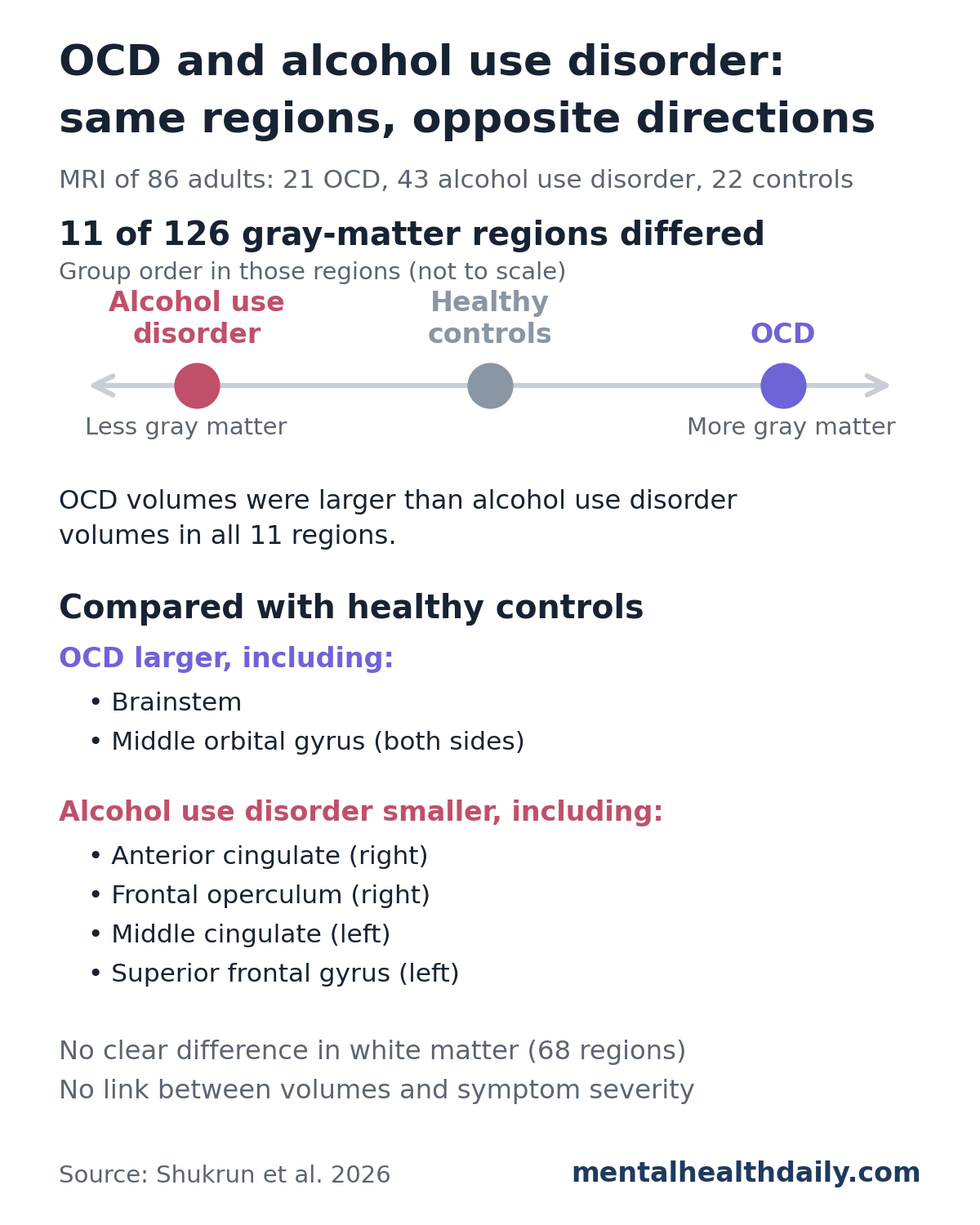
OCD vs. healthy controls: the OCD group had significantly larger volumes in regions including the brainstem (p = 0.0001) and the right and left middle orbital gyrus (p = 0.0492 and p = 0.0222).1
Alcohol use disorder vs. healthy controls: the alcohol use disorder group had significantly smaller volumes in several frontal and cingulate regions, including the right anterior cingulate (p = 0.0023), right frontal operculum (p = 0.0014), left middle cingulate (p = 0.0023), and left superior frontal gyrus (p = 0.0023).1
Many of these regions belong to the cortico-striato-thalamo-cortical (CSTC) circuit, a loop that runs from the frontal cortex through the striatum and thalamus and back. It supports self-control, habit learning, and reward, and it is the circuit most often linked to compulsive behavior.
The researchers concluded that similar compulsive behavior in the two disorders rests on different structural changes within the same circuits, rather than on one shared brain signature.1
White Matter and Symptom Severity Showed No Clear Differences
White matter: no white-matter region differed between groups. Earlier studies have reported white matter changes in both disorders, but most used diffusion imaging, which measures the integrity of nerve fibers rather than their bulk volume. The researchers noted that diffusion scans in the same alcohol cohort had shown white matter differences from controls.1
Symptom severity: gray-matter volume in the 11 regions did not correlate with OCD severity (Y-BOCS) or with alcohol craving on the Penn Alcohol Craving Scale (PACS).1 Small groups and total scores that mix different symptom types, such as contamination fears vs. checking, may have hidden a link.
How the Results Compare With Earlier OCD and Addiction Brain Studies
For alcohol use disorder, the shrinkage fits a consistent literature. A 2022 meta-analysis by Stevens et al. pooled 19 studies (736 people with alcohol use disorder, 827 controls) and found less gray matter in frontal, limbic, temporal, and cerebellar regions.3
OCD is less consistent, and earlier work has pointed in both directions:
- Shared reductions: the same Stevens et al. meta-analysis (25 OCD studies, 995 people with OCD) found reductions in frontal and insular regions, with overlap between OCD and alcohol use disorder in the anterior cingulate and insula. It also found OCD-specific increases in the hypothalamus and brainstem, which matches the brainstem result here.3
- Common frontal loss, OCD-only increase: a 2021 meta-analysis by Klugah-Brown et al. compared OCD with alcohol, cocaine, and nicotine use disorders. All showed less gray matter in the left inferior frontal gyrus, a region involved in stopping actions, and only OCD showed an increase, in the left putamen (part of the striatum), linked to more severe OCD symptoms.4
- Large OCD mega-analysis: de Wit et al. pooled raw scans from 412 people with OCD and 368 controls and found smaller frontal gray matter, including the anterior cingulate, alongside larger cerebellar gray matter.5
The 2026 study therefore agrees with earlier work on alcohol-related shrinkage and on some OCD-specific increases. It differs on the anterior cingulate: the meta-analyses found smaller volumes there in OCD, while this study found no reduction, with OCD volumes above those of the alcohol use disorder group.135
One advantage of this study is that everyone was scanned on the same scanner and processed the same way, and people with both disorders were excluded. Meta-analyses combine many different methods and often include mixed samples.1
Limits of This Small, Cross-Sectional MRI Study
This was a single snapshot of 86 people, so it can describe group differences but cannot say what caused them. The main limits:1
- Cause vs. consequence: smaller volumes in alcohol use disorder could reflect years of heavy drinking, a pre-existing vulnerability, or both. Larger volumes in OCD could reflect the illness, treatment, or compensation.
- Small, unequal groups: 21, 43, and 22 people from one center, with the controls recruited only during the alcohol study.
- Age gap: the alcohol use disorder group averaged 42.1 years, compared with 35.0 for controls and 31.5 for OCD. Age was adjusted for, but some effect may remain.
- Medications: the OCD group was a mix of unmedicated people and people taking SSRIs (selective serotonin reuptake inhibitors) alone or with other drugs, and medication histories were not available for the other groups.
- Unmeasured factors: body mass index, depression and anxiety symptom levels, illness duration, and lifetime alcohol exposure were not accounted for.
- Slightly different scan settings: the OCD scans used somewhat different MRI parameters than the alcohol and control scans, though on the same scanner.
The researchers suggest that imaging markers could eventually help with diagnosis and treatment planning. Group averages in 11 regions are a long way from that: no scan-based test was built or validated here, and the study did not report how well volumes told individual patients apart.
What the Findings Mean for Compulsive Behavior in OCD and Addiction
- Similar behavior, different brains: compulsive rituals and compulsive drinking may look alike, but in this study the underlying gray matter changed in opposite directions.
- Same circuits involved: the differences clustered in frontal, cingulate, and brainstem regions linked to self-control, habits, and reward, which fits the idea that compulsivity runs through shared circuits even when the changes differ.
- Treatment implications remain open: the finding supports disorder-specific approaches in principle, but it does not change current treatment for either condition.
- Next steps: larger studies that follow people over time and combine volume, diffusion, and functional scans could show whether these differences come before illness or develop with it.
References
- Shukrun D, Shalev H, Gulevsky T, Harel M, Zangen A. Distinct structural abnormalities in obsessive-compulsive and alcohol use disorders. European Archives of Psychiatry and Clinical Neuroscience. 2026. doi:10.1007/s00406-026-02375-y
- Everitt BJ, Robbins TW. Neural systems of reinforcement for drug addiction: from actions to habits to compulsion. Nature Neuroscience. 2005;8:1481–1489. doi:10.1038/nn1579
- Stevens BW, Morris JK, Diazgranados N, Ramchandani VA. Common gray matter reductions in alcohol use and obsessive-compulsive disorders: a meta-analysis. Biological Psychiatry: Global Open Science. 2022;2:421–431. doi:10.1016/j.bpsgos.2021.11.010
- Klugah-Brown B, Jiang C, Agoalikum E, et al. Common abnormality of gray matter integrity in substance use disorder and obsessive-compulsive disorder: a comparative voxel-based meta-analysis. Human Brain Mapping. 2021;42:3871–3886. doi:10.1002/hbm.25471
- de Wit SJ, Alonso P, Schweren L, et al. Multicenter voxel-based morphometry mega-analysis of structural brain scans in obsessive-compulsive disorder. American Journal of Psychiatry. 2014;171(3):340–349. doi:10.1176/appi.ajp.2013.13040574