
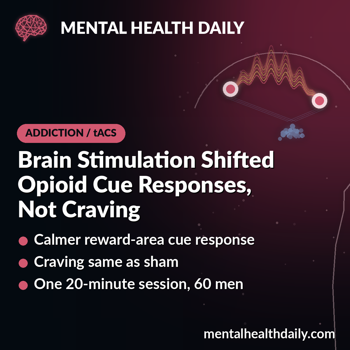
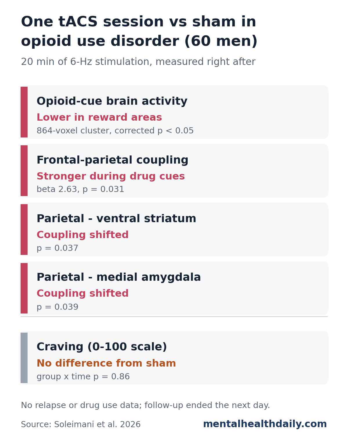
A single 20-minute session of scalp brain stimulation changed how the brains of men with opioid use disorder reacted to drug cues, but it did not change how much they craved opioids. In a randomized, sham-controlled trial of 60 men, active stimulation reduced opioid-cue activity in a cluster covering the striatum, amygdala, and ventral tegmental area, while craving ratings did not differ between the groups (group-by-time p = 0.86).1
That makes this a study about reaching the right circuits. It shows the stimulation engaged reward and control networks linked to craving. It did not test whether the method helps anyone stay off opioids.
Research Highlights
- Reward-area cue response dropped: after active stimulation, opioid-cue activity fell relative to sham in an 864-voxel cluster spanning the striatum, amygdala, ventral tegmental area, and posterior cingulate cortex (corrected for whole-brain testing).1
- Craving did not differ: craving changed over the session in both groups (p < 0.0001 for time), but the stimulation group did not differ from sham (group p = 0.81; group-by-time p = 0.86).1
- The targeted network coupled more tightly: during drug cues, the right parietal target synchronized more with the right frontal target after active stimulation (β = 2.63, p = 0.031).1
- Stronger dose, stronger network change: in the active group, a higher modeled electric field under the frontal electrode went with a larger increase in frontoparietal coupling (r = 0.43, p = 0.02); no such link appeared with sham (r = 0.08).1
- Short and narrow: 1 session, men only, early abstinence in a residential program, follow-up to the next day, and no drug use or relapse outcomes.1
What the Tulsa tACS Trial Tested
Transcranial alternating current stimulation (tACS) passes a weak electrical current that oscillates at a set rhythm through electrodes on the scalp. The idea is to nudge brain regions into firing in step. This trial used 6 Hz, which falls in the theta band (roughly 4 to 8 cycles per second), a rhythm linked to communication between the frontal and parietal parts of the brain’s executive control network.1
The researchers aimed 2 sets of high-definition electrodes at 2 spots on the right side of the head: the dorsolateral prefrontal cortex (toward the front of the head, at the EEG position F4) and the inferior parietal cortex (farther back, at P4). Both sites were stimulated in phase, so the frontal and parietal targets received the same 6-Hz rhythm at the same moment. The plan was to strengthen this control network and, through it, dampen the reward and emotion areas that light up when someone with addiction sees drug cues.1
Sixty men with opioid use disorder, average age about 34, were randomized 30 per group. All were in early abstinence at a residential addiction treatment center in Tulsa, Oklahoma, and recruitment ran from 2019 to 2023.1 The 2 groups were:
- Active tACS: 20 minutes of 6-Hz stimulation at 2 mA peak-to-peak over both targets.
- Sham: sham stimulation is a fake version meant to feel real. Here the device ramped the current up and down for 30 seconds at the start and end to mimic the tingling, and delivered nothing in between.
The trial was preregistered (NCT03907644) and triple-blind, and the data analysts worked with coded group labels until the preregistered analyses were done. Everyone completed both scanning sessions.1
Right before and right after stimulation, each man lay in an fMRI scanner (functional magnetic resonance imaging, which tracks blood-oxygen changes as a proxy for brain activity) and viewed alternating blocks of opioid-related and neutral images. He rated his craving from 0 to 100 after each block, and craving was also rated at baseline, around the scans, and the next day.1
Active Stimulation Quieted the Brain’s Response to Opioid Cues
The preregistered primary outcome was the change in cue-related brain activity: the response to opioid images minus the response to neutral images, before vs after stimulation, active vs sham.
The largest significant cluster covered 864 voxels across both sides of the striatum, the amygdala, the ventral tegmental area (the midbrain source of much of the brain’s dopamine), and the posterior cingulate cortex. In that cluster, the opioid-cue response fell after active stimulation compared with sham.1 These are core reward and emotion regions, and the striatum is a key hub of the brain’s reward system.
That matters because a drop in cue reactivity in these regions is a pattern seen with effective addiction treatment. A 2025 meta-analysis of 51 fMRI cue-reactivity studies with 1,787 participants found that activity in the cingulate, caudate, nucleus accumbens, and insula fell after treatment, and striatal reductions were more pronounced with active treatment than with placebo.2 The tACS result fits that pattern, although here it was measured minutes after 1 session rather than after weeks of treatment.
Craving Did Not Differ From Sham
Craving was a secondary outcome, and it did not separate the groups.
- Over time: craving shifted significantly across the 6 rating points, rising after the first round of drug images and falling afterward (F = 14.21, p < 0.0001).
- Between groups: no difference between active and sham (F = 0.056, p = 0.81).
- Group by time: no difference in how craving changed (F = 0.386, p = 0.86). Scores on the Desires for Drug Questionnaire showed the same null interaction.1
Both groups also reported lower craving the next day than at baseline, again with no difference between them. The authors note that several other single-session electrical stimulation studies have likewise found no difference in self-reported craving between active and sham groups.1
Frontoparietal Coupling Rose and Its Links to Reward Areas Shifted
The researchers also measured task-based connectivity: how closely activity in one region tracked another specifically while the men viewed drug cues rather than neutral images. Starting from the right parietal target, they found 3 significant changes in the active group relative to sham:1
- Stronger coupling within the control network. The right parietal site synchronized more with the right frontal site (β = 2.63) and with the left frontal and left parietal regions (cluster p = 0.031). This is the network the stimulation was designed to engage.
- Parietal cortex and right ventral striatum. Coupling shifted toward a more negative, opposing relationship (cluster β = 1.99, p = 0.037).
- Parietal cortex and left medial amygdala. The same kind of shift (β = 1.97, p = 0.039).
The authors read the negative shift as stronger top-down control over reward and emotion areas. Their own limitations section adds that this connectivity method measures context-dependent coupling and cannot show which region drives which.1 Resting-state connectivity, measured without drug cues, showed no significant changes.
Two exploratory analyses linked these network effects to craving and to stimulation strength. In the active group, men whose frontoparietal coupling rose more tended to show a larger drop in cue-induced craving after stimulation (r = −0.37, p = 0.05). And a stronger modeled electric field under the frontal electrode went with a larger coupling increase (r = 0.43, p = 0.02), which the authors describe as preliminary evidence of a dose-response effect.1 The craving correlation sits right at the significance threshold and comes from 30 people, so it is a lead for future trials to check.
Why One Session Rarely Moves Craving
The null craving result is what earlier stimulation research would predict.
- More sessions, bigger effects: a meta-analysis of 48 studies with 1,095 participants found that stimulation of the dorsolateral prefrontal cortex had a small effect on craving and a medium effect on consumption compared with sham. Multi-session protocols beat single sessions, with a linear relationship between the number of sessions and craving reduction.3
- Clinical trials use courses, not single doses: a 2024 systematic review of 94 neuromodulation studies in substance use disorders found repetitive transcranial magnetic stimulation (rTMS) reduced substance use and craving with medium-to-large effect sizes, with the best results from multiple sessions over the left dorsolateral prefrontal cortex. Transcranial direct current stimulation produced medium but more variable effects.4
Those reviews measured craving and substance use directly. The Tulsa trial measured brain activity after 1 session. The two kinds of evidence answer different questions, and this trial answers the earlier one: whether this particular montage reaches the circuits it was built to reach. In this sample, it did.
Craving itself is worth targeting. A 2022 meta-analysis of 237 studies and 51,788 participants found that drug cues and craving prospectively predicted later drug use and relapse; in real-time phone-based studies, cues and craving went with about double the odds of subsequent use (odds ratio 2.05).5 Shrinking cue reactivity is therefore a reasonable path to test, but the payoff has to show up in craving and use, and that requires longer trials.
Where This Fits Next to Established Opioid Use Disorder Treatment
Opioid use disorder already has effective treatment. Medications such as methadone and buprenorphine are the standard of care, and staying on them is linked to much lower death rates: in a meta-analysis of cohort studies, all-cause mortality was 11.3 per 1,000 person-years during methadone treatment vs 36.1 out of it, and 4.3 vs 9.5 for buprenorphine.6 Brain stimulation for opioid use disorder is an experimental add-on idea. The trial report does not say whether participants were taking these medications.
Limitations
- 1 session, short follow-up: outcomes ended the day after stimulation. There were no drug use, relapse, or urine toxicology outcomes.
- Men only: the residential center had very few women, and the authors call this a significant limitation given evidence for sex differences in stimulation response.
- Narrow setting: early abstinence in residential treatment; outpatients and people still using may respond differently.
- Standard targeting: electrodes sat at fixed EEG positions rather than on each person’s own cue-reactive network. In the authors’ earlier methamphetamine work, the parietal region best connected to F4 sat slightly off P4.
- Technical details: 2 identical-model MRI scanners were used, and the cue task included an attention check that slowed responses during drug-cue blocks.
Blinding held up well: 24 of 30 sham participants and 25 of 30 active participants guessed they had received real stimulation right after the post-stimulation scan. Only 2 severe side effects were reported, trouble concentrating in 1 sham participant and skin redness the next day in 1 active participant.1
Practical Takeaways
- A mechanism result: 1 session of theta-band frontoparietal tACS changed cue-related activity and coupling in reward and control circuits in men with opioid use disorder.
- No craving benefit yet: craving did not differ from sham, and relapse was not studied.
- Not a treatment option now: tACS for opioid use disorder is experimental. Medications for opioid use disorder remain the evidence-based core of care.
- What comes next: the authors call for multi-session trials with brain-imaging-guided targeting and long-term follow-up with objective drug use measures.
References
- Soleimani G, Kuplicki R, Paulus MP, Ekhtiari H. Targeting cortico-striatal-amygdalar networks via theta-band frontoparietal synchronization in opioid use disorder: a randomized tACS-fMRI trial. Molecular Psychiatry. 2026;31:6056–6068. doi:10.1038/s41380-026-03694-1
- Evohr B, Wenzel S, Chen N, et al. Cue-elicited brain activity and treatment outcomes in substance use disorders: a meta-analysis. JAMA Network Open. 2025;8(12):e2548809. doi:10.1001/jamanetworkopen.2025.48809
- Song S, Zilverstand A, Gui W, Li HJ, Zhou X. Effects of single-session versus multi-session non-invasive brain stimulation on craving and consumption in individuals with drug addiction, eating disorders or obesity: a meta-analysis. Brain Stimulation. 2019;12(3):606–618. doi:10.1016/j.brs.2018.12.975
- Mehta DD, Praecht A, Ward HB, et al. A systematic review and meta-analysis of neuromodulation therapies for substance use disorders. Neuropsychopharmacology. 2024;49:649–680. doi:10.1038/s41386-023-01776-0
- Vafaie N, Kober H. Association of drug cues and craving with drug use and relapse: a systematic review and meta-analysis. JAMA Psychiatry. 2022;79(7):641–650. doi:10.1001/jamapsychiatry.2022.1240
- Sordo L, Barrio G, Bravo MJ, et al. Mortality risk during and after opioid substitution treatment: systematic review and meta-analysis of cohort studies. BMJ. 2017;357:j1550. doi:10.1136/bmj.j1550