
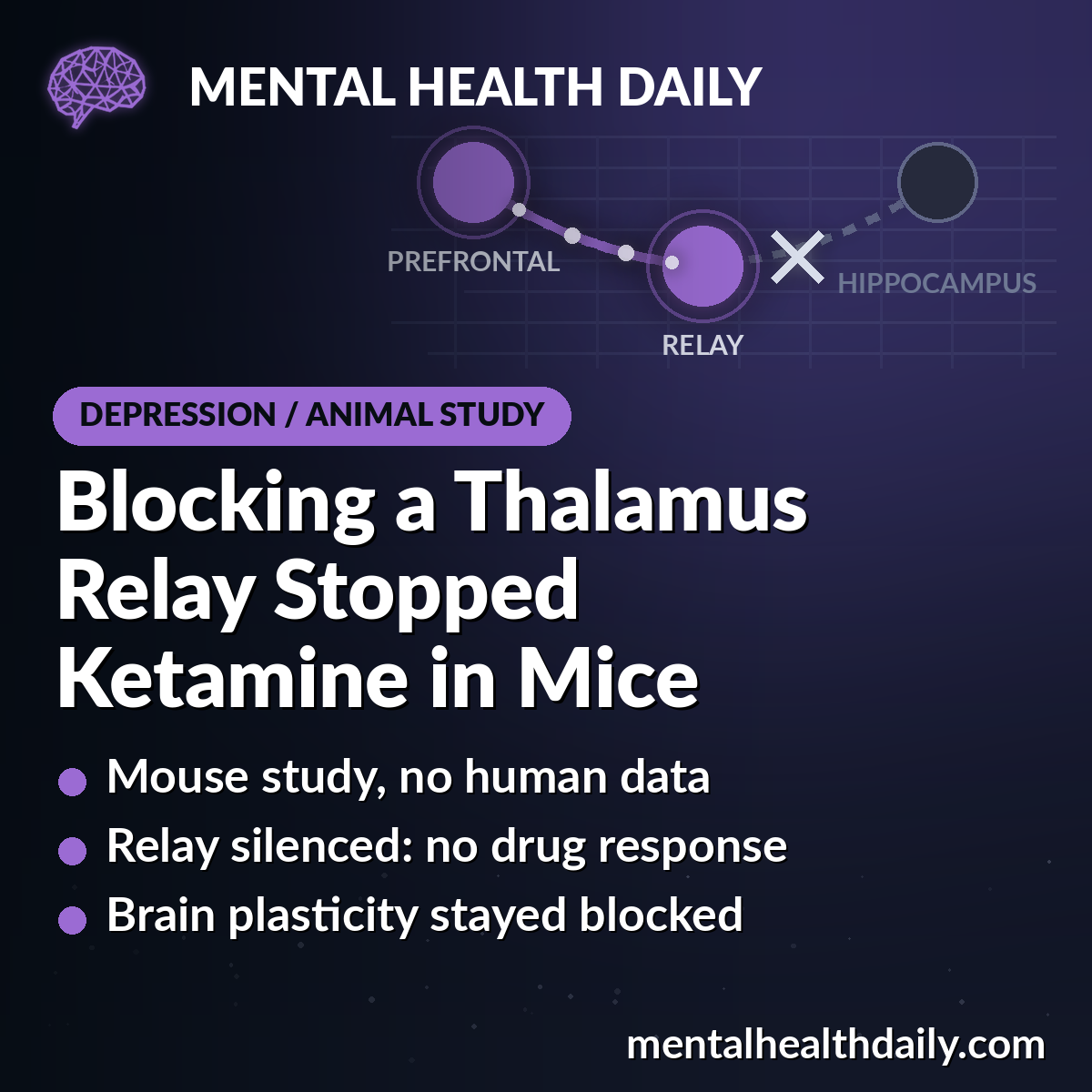
Ketamine stopped working in stressed mice when researchers switched off a small relay station in the thalamus, or either end of the prefrontal-to-hippocampus pathway running through it, according to a 2026 mouse study in Nature Communications.1
Research Highlights
- Relay silenced, drug response gone: a single 10 mg/kg dose of ketamine reduced swim immobility in stressed mice with the circuit left intact (7 mice, P = 0.0093), but changed nothing when the nucleus reuniens was switched off (8 mice, P = 0.6987).1
- Cutting either end did the same: blocking prefrontal fibers going into the relay (8 mice, P = 0.5380) or relay fibers going out to the hippocampus (5 mice, P = 0.6726) removed the same response.1
- Plasticity blocked too: ketamine normally restored long-term potentiation, a lasting strengthening of synapses, in hippocampal slices from stressed mice; cutting the prefrontal-to-relay link prevented that rescue (9 cells per group, P = 0.0264).1
- Turning the prefrontal node on worked like a treatment: switching on excitatory neurons in the infralimbic cortex overnight reduced immobility (12 mice, P = 0.0003) and restored hippocampal plasticity (14 cells, P = 0.0023).1
- Mouse mechanism work: no people were studied, group sizes were 4 to 17 mice, and the silencing method has no clinical equivalent. It explains how an antidepressant response is wired, not how to treat anyone.1
Two Theories of Depression That Rarely Meet
Depression research runs on two big ideas. One says symptoms come from faulty communication between brain regions. The other says the brain loses some of its ability to rewire itself — its neuroplasticity.1
Maxime Veleanu et al., at the University of Freiburg and the University of Strasbourg, set out to connect the two in mice: find a circuit whose activity decides whether hippocampal synapses can still change.1
The three regions involved:
- Infralimbic cortex (IL): a lower part of the medial prefrontal cortex, the front-of-brain area that regulates emotion. Its closest human counterpart is the subcallosal cingulate cortex, the region targeted by deep brain stimulation for treatment-resistant depression.
- Nucleus reuniens (RE): a thin nucleus in the midline thalamus. The prefrontal cortex has almost no direct excitatory wiring to the hippocampus, so this nucleus carries the signal — a junction box between the two.
- Ventral hippocampus (vHIPP): the front-lower end of the hippocampus, linked to emotion and stress rather than to the spatial memory handled by its dorsal end.
How Researchers Switched Single Brain Circuits On and Off in Mice
Mice went through a 5-day stress procedure that produces lasting depression-like behavior: less interest in sugar water, less exploring, and faster loss of active struggling in swim and tail-suspension tests.1
Chemogenetics was the main tool. A harmless virus delivers an engineered receptor into chosen neurons, and those neurons — and only those — respond to a drug that does nothing elsewhere in the brain.1
- hM3Dq: the on-switch version, used to activate infralimbic neurons.
- hM4Di: the off-switch version, used to silence the reuniens or one specific projection.
- mCherry: a fluorescent protein with no switch, used as the control virus.
Single projections were targeted by injecting a virus that travels backward along axons into the destination region, so that only neurons sending fibers there carried the off-switch.1
Investigators were blinded to treatment group until data collection, mice were randomly assigned, and males and females were included in equal numbers.1
Prefrontal Activation Reversed Depression-Like Behavior
Overnight activation of infralimbic excitatory neurons reversed the stress effect across several tests: swim immobility (12 mice, P = 0.0003), tail-suspension immobility (P = 0.0007), sugar-water preference (P = 0.0068), novel-object exploration (P = 0.0027) and social preference (P = 0.0415).1
Distance traveled in an open field (P = 0.3634) and general exploring in the home cage were unchanged, so the behavior changes were not simply mice becoming more active.1
Overnight, not acute: a single acute dose of the activating drug did nothing in the swim or tail-suspension tests. Only overnight activation worked.1
One subregion only: activating the prelimbic cortex, the neighboring prefrontal subregion just above the infralimbic, left immobility and sugar-water preference unchanged.1
That specificity is consistent with earlier rodent work. Fuchikami et al. reported in 2015 that silencing the infralimbic cortex blocked ketamine's antidepressant-like and anxiety-reducing effects in rats, that ketamine infused directly into it reproduced them, and that optogenetic stimulation of infralimbic but not prelimbic neurons recapitulated them.2
Hippocampal Plasticity Came Back With Prefrontal Stimulation
Long-term potentiation (LTP) is the lasting strengthening of a synapse after it is used in a particular pattern — the standard laboratory measure of the brain's capacity to rewire. Researchers measured it in brain slices, by pairing incoming signals with the receiving cell's own firing.1
In slices from unstressed mice, synapses strengthened and stayed strengthened. In slices from stressed mice, this strengthening was absent (10 vs. 11 recorded cells, P = 0.0135).1
Overnight infralimbic activation brought it back (7 vs. 14 cells, P = 0.0023). Acute activation did not.1
New thalamic synapses appeared: markers of incoming thalamic connections (VGLUT2) increased in the ventral hippocampus (P = 0.0081) but not the dorsal hippocampus (P = 0.0809), and markers of local and inhibitory connections did not change.1
Plasticity-related genes followed the same split: several switched on in the ventral hippocampus and none in the dorsal hippocampus.1
Silencing the Thalamic Relay Removed Ketamine's Effect
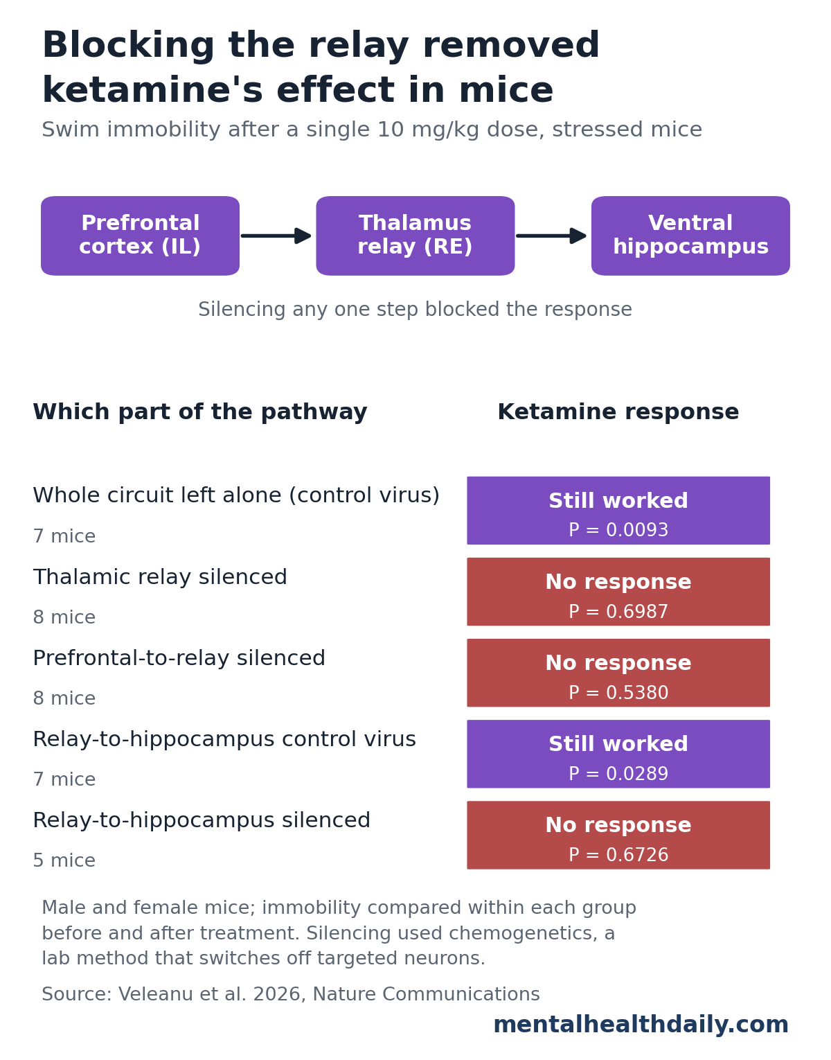
Ketamine 10 mg/kg reduced swim immobility in stressed mice carrying the control virus (7 mice, P = 0.0093). With the reuniens silenced, the same dose changed nothing (8 mice, P = 0.6987).1
Silencing only the prefrontal fibers entering the relay produced the same loss of response (8 mice, P = 0.5380), as did silencing only the fibers leaving the relay for the ventral hippocampus (5 mice, P = 0.6726).1
The plasticity results matched the behavior. Ketamine restored hippocampal LTP in slices from stressed mice, and blocking the prefrontal-to-relay link prevented that restoration (9 cells per group, P = 0.0264).1
Silencing the relay also canceled the effects of prefrontal activation itself — behavior and plasticity alike — while silencing the relay on its own left immobility unchanged, so the nucleus was not simply driving behavior by itself.1
What Blocking a Relay Can and Cannot Show
Ketamine acts on receptors throughout the brain, including directly in the hippocampus, so a thalamic relay being necessary is not the same as it being where the drug works.1
Veleanu et al. offered two explanations. Ketamine may act locally at first, with the effect lasting only while a wider prefrontal–thalamus–hippocampus loop stays engaged. Or it may act at several points, including the prefrontal cortex, and need the loop to carry the effect forward.1
The second reading fits earlier rat work: Carreno et al. found in 2016 that inactivating the ventral hippocampus prevented ketamine's sustained response, that activating the ventral-hippocampus-to-prefrontal pathway mimicked ketamine, and that inactivating it reversed the response.3
Together those results describe a loop rather than a one-way line: prefrontal cortex to thalamus to hippocampus, and hippocampus back to prefrontal cortex.
The Human Parallel Stops at Anatomy
In people with treatment-resistant depression, deep brain stimulation targets the subcallosal cingulate cortex, the region corresponding to the mouse infralimbic cortex.1
Riva-Posse et al. mapped white matter tracts in 16 such patients and found that everyone who responded — 6 at 6 months and 12 at 2 years — shared bilateral connections from the stimulated volume to medial frontal cortex, cingulate cortex and subcortical nuclei, while non-responders did not consistently show them.4
That is an anatomical overlap, not confirmation. Nobody has tested in a person whether the midline thalamus is required for ketamine to work.
Limitations of This Mouse Circuit Study
This is mechanism research in animals. Mice do not have depression; they have behavior under stress that responds to drugs that treat depression in people.
- Mice only: no human data were collected.
- Small groups: 4 to 17 mice per behavioral group and 6 to 14 recorded cells per slice group, with some single-timepoint comparisons.
- Sex differences untested: males and females were included in equal numbers and an exploratory analysis found no differences, but the study was not powered to detect them.
- Artificial silencing: chemogenetics shuts off a defined group of neurons far more completely than any available clinical procedure.
- Necessity, not location: removing a response by blocking a relay does not pin down where ketamine acts.
- All-or-nothing plasticity measure: LTP in these slices was either present or absent, which shows the switch but not how much plasticity changed.
- Industry ties: 3 of the authors report lecture fees or advisory honoraria from Johnson & Johnson, which makes esketamine.
No treatment implication: nothing here changes how ketamine or brain stimulation is used. The practical value is a target for future work — a midline thalamic nucleus that had barely been examined in antidepressant research.
References
- Veleanu M, Schuberth L, Kilias A, et al. The mPFC-reuniens-hippocampus pathway links brain circuitry and neural plasticity in antidepressant response. Nature Communications. 2026;17:9361. doi:10.1038/s41467-026-77248-y
- Fuchikami M, Thomas A, Liu R, et al. Optogenetic stimulation of infralimbic PFC reproduces ketamine's rapid and sustained antidepressant actions. Proceedings of the National Academy of Sciences. 2015;112(26):8106–8111. doi:10.1073/pnas.1414728112
- Carreno FR, Donegan JJ, Boley AM, et al. Activation of a ventral hippocampus–medial prefrontal cortex pathway is both necessary and sufficient for an antidepressant response to ketamine. Molecular Psychiatry. 2016;21:1298–1308. doi:10.1038/mp.2015.176
- Riva-Posse P, Choi KS, Holtzheimer PE, et al. Defining critical white matter pathways mediating successful subcallosal cingulate deep brain stimulation for treatment-resistant depression. Biological Psychiatry. 2014;76(12):963–969. doi:10.1016/j.biopsych.2014.03.029