Autistic children had more complex, irregular brain-wave signals at rest than typically developing children in a 2026 EEG study of 186 kids aged 3 to 11 in Beijing.1
The gap was widest in preschoolers aged 3 to 6 and smaller, and limited to some brain regions, in school-age children.
Research Highlights
- Higher complexity in autism: across all 186 children, the 80 autistic kids had higher multiscale entropy, a measure of signal complexity, in all 6 brain regions tested (all p < 0.001).1
- Biggest gap at ages 3 to 6: among 60 autistic and 76 typically developing preschoolers, the difference showed up in every region and across many signal timescales.1
- Narrower gap at ages 6 to 11: among 20 autistic and 30 typically developing school-age children, the difference was significant in 4 of 6 regions, mainly at the fastest timescales and over the front of the head.1
- Different age patterns: complexity was higher in older typically developing children at every timescale, while the age pattern in autistic children was patchy, rising in some regions and falling in others.1
- Snapshot, not a growth curve: each child was recorded once, so the age comparisons are between different children rather than tracked development.1
What “Neural Signal Complexity” Means on an EEG
Electroencephalography (EEG) records the brain’s electrical activity through sensors on the scalp. The result is a set of wavy lines, one per sensor, that rise and fall many times per second.
Neural signal complexity describes how predictable those wavy lines are. A signal that keeps repeating the same short patterns is simple. A signal whose next wiggle is hard to guess from the last few is more complex.
Researchers measure this with entropy, a number that rises as a signal becomes less predictable. This study used multiscale entropy (MSE), which checks predictability at many speeds:
- Fine timescales: the raw signal, capturing fast moment-to-moment fluctuations
- Coarse timescales: the same signal averaged into longer and longer chunks (up to 20 steps here), capturing slower patterns
Higher complexity is not automatically better. In healthy development, complexity tends to rise as brain networks mature. Very high complexity can also mean noisier, less coordinated signaling, and the researchers read the extra complexity in autistic children that way.1
Who Was Studied and How
Han et al. recorded about 5 minutes of eyes-open, resting EEG from each child using a 128-sensor head net.1
- Autistic group: 80 children diagnosed by DSM-IV criteria (65 boys, 15 girls; average age 5.7)
- Comparison group: 106 typically developing children from Beijing kindergartens and primary schools (79 boys, 27 girls; average age 5.9), matched on age and sex
- Excluded: Rett syndrome, global developmental delay, and other neurological or psychiatric disorders
- Analysis: MSE at 20 timescales, averaged over 6 brain regions (frontal, central, parietal, occipital, left temporal, right temporal), with age and sex taken into account
Both groups kept a similar number of clean EEG segments after noisy stretches were removed. That makes it less likely that fidgeting or movement artifacts produced the group difference.1
Autistic Children Had Higher EEG Complexity in All 6 Brain Regions
Across the full sample, autistic children had higher multiscale entropy than typically developing children in every region (all p < 0.001). Sensor-by-sensor comparisons found the same pattern across nearly all sensors and timescales.1
The size of the difference also depended on timescale in every region, so the gap was not uniform from fast to slow signal patterns. Sex had no significant effect in any region.1
The Complexity Gap Was Smaller in School-Age Children
To look at age, the researchers split the children into 2 groups.1
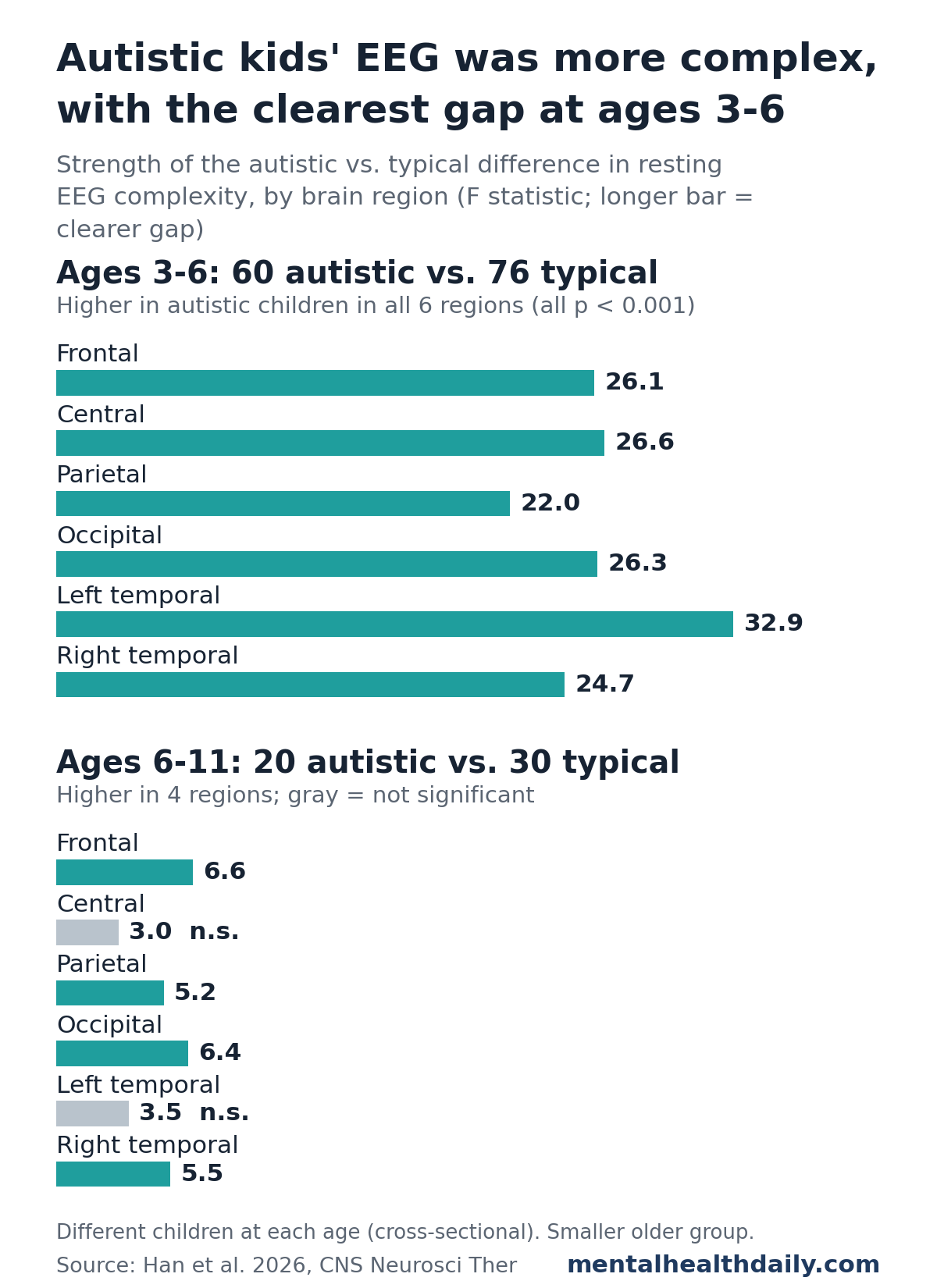
- Preschool, ages 3 to 6 (60 autistic, 76 typical): higher complexity in autistic children in all 6 regions and across all timescales
- School age, ages 6 to 11 (20 autistic, 30 typical): higher complexity in the frontal, parietal, occipital and right temporal regions, but not the central or left temporal regions
In the older group, the differences that held up were mostly at the fastest timescales, and scalp maps placed them mainly over the frontal region, which supports planning, attention and social thinking.1
Sample size caveat: the school-age comparison involved only 50 children, compared with 136 in the preschool comparison. Smaller groups make real differences harder to detect, so some of the shrinking gap may reflect fewer children rather than less difference.
EEG Complexity Rose With Age in Typical Children but Not Evenly in Autistic Children
In typically developing children, older kids had higher complexity at every timescale, with the strongest age link at the slower timescales (steps 11 to 20). That fits the idea that brain signals grow richer as networks mature.1
In autistic children, the age pattern was uneven:1
- Occipital region (back of the head, vision): complexity was higher in older children, mainly at fast timescales (steps 1 to 5)
- Left and right temporal regions (sides of the head, hearing and language): complexity was lower in older children, mainly at middle timescales (steps 6 to 15)
One reading is that autistic children start with high complexity and older autistic children look more like their typical peers. The researchers also raise compensation by higher-order brain systems. Because each child was recorded only once, the study cannot tell whether any individual child’s brain signals actually changed with age.1
Links Between EEG Complexity and Autism Symptoms Were Mixed
The researchers compared complexity with parent-reported symptom scores on the Autism Behavior Checklist (ABC) and the Social Responsiveness Scale (SRS), where higher scores mean more autism-related difficulties.1
- ABC: complexity in frontal and parietal regions was significantly linked to sensory, social interaction and language scores
- SRS social cognition: complexity in central and parietal regions was positively correlated with this score
The direction is hard to pin down. The results section says higher complexity may go with better social thinking, while the discussion says higher frontal and parietal complexity went with more difficulty in sensory processing, communication, language and social cognition. Correlation strengths were not reported, and many sensor-and-timescale pairs were tested, so these links are exploratory.1
Earlier MEG and EEG Studies Found Similar Age Effects, With Exceptions
Younger autistic children, more variable signals. A 2016 study by Takahashi et al. used magnetoencephalography (MEG), which records the brain’s magnetic fields, in 43 autistic and 72 typically developing young children watching silent videos. Signal variability rose with age in typical children, followed an atypical age pattern in autistic children, and was higher in autistic children, especially the youngest.2
Infants with an autistic older sibling. A 2011 study by Bosl et al. found that resting EEG complexity followed a different path from 6 to 24 months in infants at higher familial likelihood of autism. The differences were greatest at 9 to 12 months.3
Adults during a task: lower complexity. A 2011 study by Catarino et al. found reduced EEG complexity over temporo-parietal and occipital regions in 15 autistic adults compared with 15 controls while they matched pictures of faces and chairs.4
Taken together, the direction of the difference seems to depend on age, recording conditions (rest versus a task) and method. The 2026 study adds a fairly large sample of young children at rest.
Limits of This Cross-Sectional EEG Study
- One recording per child. Age patterns compare younger and older children, not the same children over time. Long-term follow-up would be needed to show real developmental change.
- Small school-age group. Only 20 autistic and 30 typically developing children were aged 6 to 11.
- Many comparisons. The analysis covered 6 regions, 20 timescales and dozens of sensors. Group comparisons were corrected for this, but a correction for the symptom correlations was not described.
- Missing context. No data on IQ or developmental level, medication or therapy were reported, and the sample was mostly boys from one city.
- Indirect measure. Multiscale entropy describes signal patterns, not the cell-level processes behind them, and resting EEG says nothing about how complexity shifts during social or attention tasks.
The findings describe group averages. EEG complexity is not a test for diagnosing autism in an individual child, and the study offers no treatment implications.
References
- Han J, Liang L, Song R, et al. Atypical age-related patterns of neural signal complexity in autism spectrum disorder. CNS Neuroscience & Therapeutics. 2026;32:e71173. doi:10.1002/cns.71173
- Takahashi T, Yoshimura Y, Hiraishi H, et al. Enhanced brain signal variability in children with autism spectrum disorder during early childhood. Human Brain Mapping. 2016;37(3):1038–1050. doi:10.1002/hbm.23089
- Bosl W, Tierney A, Tager-Flusberg H, Nelson C. EEG complexity as a biomarker for autism spectrum disorder risk. BMC Medicine. 2011;9:18. doi:10.1186/1741-7015-9-18
- Catarino A, Churches O, Baron-Cohen S, Andrade A, Ring H. Atypical EEG complexity in autism spectrum conditions: a multiscale entropy analysis. Clinical Neurophysiology. 2011;122(12):2375–2383. doi:10.1016/j.clinph.2011.05.004