
Neurons that tracked a remembered smell turned up all over the mouse brain in a newly released dataset of 33,028 neurons recorded from 40 mice. The 2026 data paper makes those recordings free for other scientists to study how working memory works.1
Research Highlights
- Memory neurons in every well-sampled region: in all 34 brain regions with more than 100 recorded neurons, between 16% and 67% fired differently during the memory delay depending on which odor the mouse was holding in mind. Across those 34 regions, that came to 7,568 of 24,667 neurons, or about 31%.1
- Highest in smell and frontal areas: the largest shares were in olfactory regions (67% in the taenia tecta, 59% in the anterior olfactory nucleus) and prefrontal areas (48% infralimbic, 44% orbital). Visual, touch, motor and retrosplenial cortex sat at 16% to 18%.1
- A large, documented recording set: 116 sessions and 223 Neuropixels probe insertions covered 62 brain regions, with about 17 GB of spike data released in a standard format.1
- Built for questions about replay: the files include the quiet 10-second gaps between trials, so researchers can look for memory-related activity patterns that repeat when the mouse is not doing the task.12
- Mouse data only: this is a resource paper in trained male mice. It says nothing directly about human memory, memory problems or treatment.1
What Working Memory Is and How the Mice Were Tested
Working memory is the brain’s short-term scratchpad: it holds a piece of information for a few seconds so you can use it, like keeping a phone number in mind while you dial. Working memory problems are common in conditions such as schizophrenia, which is one reason neuroscientists want to understand how the brain does it.7
Since the 1970s, researchers have known that some neurons keep firing during a memory delay. In 1971, Fuster and Alexander recorded cells in the monkey prefrontal cortex and thalamus that changed their firing during the delay of a memory task, and some kept firing at a higher rate through the whole wait.3
Ermeng Huang et al. trained head-fixed mice on an olfactory delayed paired-association task. Each trial ran like this:1
- Sample: one of 2 odors puffed for 1 second.
- Delay: a wait of 3 or 6 seconds with no odor. The mouse has to hold the first odor in memory.
- Test: a second odor for 1 second.
- Response: if the 2 odors formed a learned pair, licking in the next second earned a drop of water.
A 10-second break followed each trial, and a typical session had about 240 trials. Recording started only after a mouse got at least 75% of 40 trials in a row correct. The researchers alternated the 3- and 6-second delays to loosen the link between holding the memory and expecting a reward.1
33,028 Neurons Recorded With Neuropixels Probes
Neuropixels probes are thin silicon needles, here 10 mm long, with hundreds of recording sites along the shaft. One insertion can pick up the electrical spikes of many individual neurons across several brain structures at once.1
Software called Kilosort2 sorted the raw signals into individual neurons, keeping only units that passed fixed quality limits. After the experiments, the team traced each probe’s path through brain slices and matched it to the Allen Mouse Brain Common Coordinate Framework, a standard 3D map of the mouse brain.1
- Mice: 40 adult male C57BL/6J mice
- Recordings: 116 sessions, 223 probe insertions, collected in 2019 and 2020
- Neurons: 33,028 curated single units in 62 regions; 34 regions had more than 100 neurons each
- Brain location: 25,347 neurons have a fine-grained region label, 1,742 have only a broader label, and 5,939 have no region label, mostly because their probe track could not be reconstructed with confidence
Each session is a Neurodata Without Borders file, a common format for brain recordings that both Python and MATLAB can read. The files hold spike times, quality scores, brain coordinates, trial events and ready-made firing-rate summaries, plus demonstration scripts for common analyses.1
Odor-Memory Neurons Were Found in All 34 Well-Sampled Regions
The researchers flagged a neuron as memory-content-selective when its firing during the delay differed significantly between the 2 sample odors. Its activity carried information about which odor the mouse was remembering.1
Every one of the 34 regions with more than 100 recorded neurons had a sizable share of these cells. Smell-processing regions and the prefrontal cortex led, while visual, touch and motor areas had the smallest shares.1
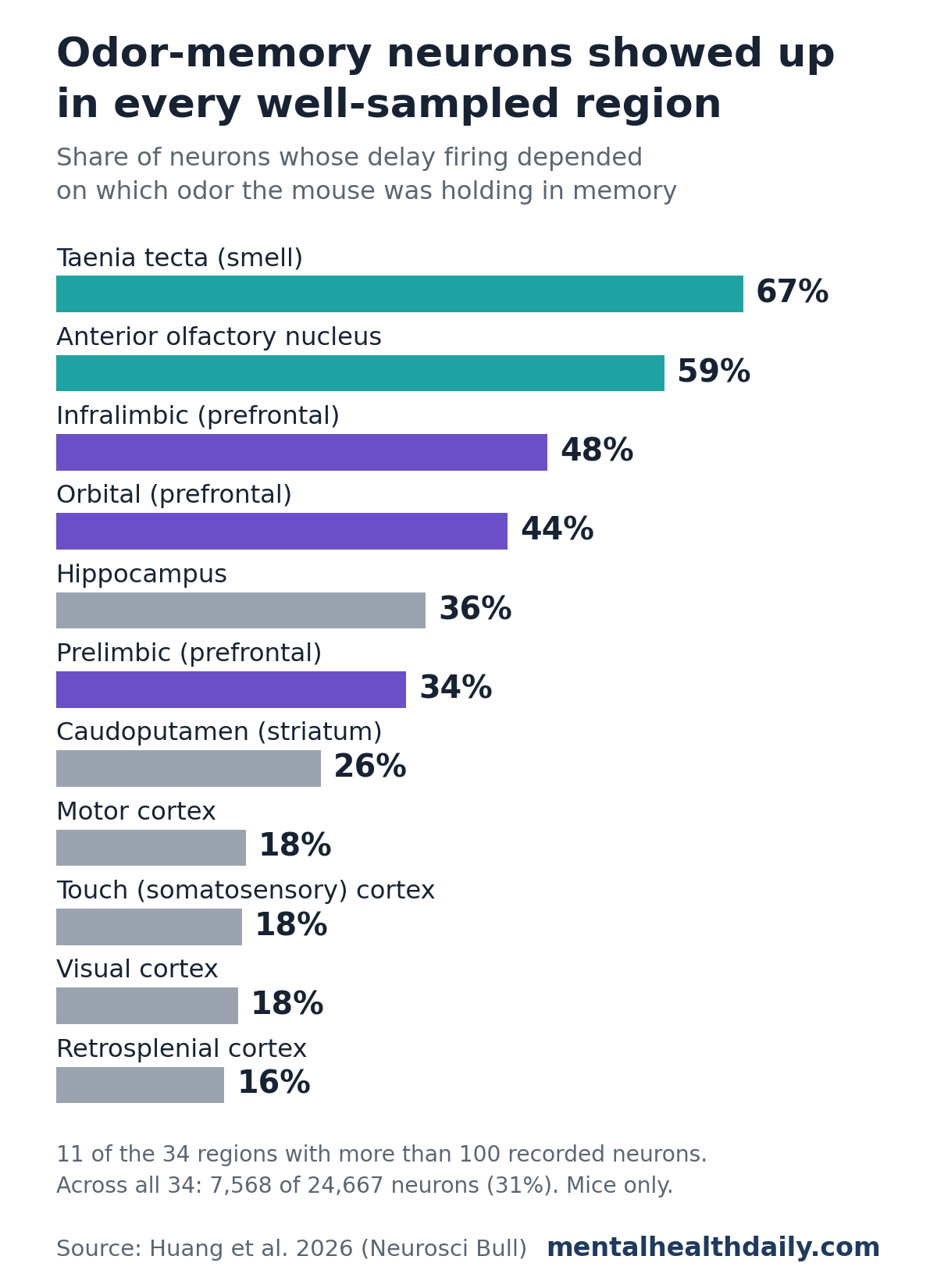
Large regions carried big counts: the orbital area alone held 1,091 selective neurons, the caudoputamen (part of the striatum, involved in movement and reward learning) held 902 and the hippocampus held 670.1
Reading the smell-area numbers: the remembered item was an odor, so olfactory regions would be expected to rank high. The team measured that the odor itself cleared to under 1% of its peak within 1 second of the valve closing, so the delay activity was not simply the smell still hanging in the air.1
Some of the delay firing in smell areas could still be lingering sensory processing inside the neurons rather than memory in the strict sense. The dataset gives researchers a way to test that question.
These percentages are descriptive counts from the dataset, not a formal comparison between regions. Some neurons in any region will pass a significance test by chance, so the lowest shares are less informative than the overall spread.
Earlier Brain-Wide Mouse Recordings Also Found Spread-Out Signals
The idea that working memory lives in one spot has faded. A 2017 review of human and monkey studies found no single storage site: activity carrying remembered information appears in sensory, parietal and prefrontal cortex.4
Large Neuropixels surveys in mice point the same way for other behaviors:
- Steinmetz et al. 2019: about 30,000 neurons in 42 regions during a visual choice task. Neurons in nearly every region responded when the mouse started to move, while signals about the choice itself were rare.5
- International Brain Laboratory 2025: 621,733 neurons from 139 mice across 12 labs, all running the same decision task. Activity linked to upcoming movement and to reward was found almost everywhere in the brain, and the data were made public.6
Those decision tasks had no multi-second memory delay. The new dataset fills that gap: a brain-wide recording set built around holding information in mind for 3 to 6 seconds.1
What Researchers Can Do With the Dataset
The same recordings were the basis for a 2025 study by the same group. It found that neurons coupled their spikes within milliseconds, both within and across regions, and that this coupling was concentrated among memory neurons that preferred the same odor.2
Chains of these coupled spikes formed repeating activity patterns. Those patterns replayed before and after the task and during the breaks between trials, and they were organized in layers, with larger patterns lasting longer and involving more neurons.2
With the data public, other groups can test those findings and ask new questions:1
- Theory testing: check competing models of how brief spikes add up to seconds of memory, such as sustained firing in recurrent networks or chains of neurons firing in sequence
- Replay: look for memory-related activity that repeats when the mouse is resting between trials
- Errors: compare correct trials with mistakes, since incorrect trials are kept in the files
- Brain-inspired AI: use layered spike patterns as a guide for artificial network designs
Limitations of This Mouse Working Memory Dataset
- Animal data: all 40 mice were young adult males of one lab strain, so sex differences and other strains are not covered.
- One task, one sense: the mice remembered odors in a 2-odor task. Memory for places, sounds or sequences may use different regions in different proportions.
- Trained animals only: recording began after the mice learned the task, so the data cannot show how working memory develops during learning.
- Uneven coverage: 28 of the 62 regions had fewer than 100 neurons, and 5,939 neurons have no region label.
- Automated sorting: neurons were selected by software rules without manual review, and the 1-spike-per-second minimum leaves out quietly firing cells (a supplementary file includes some of them).
- Description, not a new test: the paper documents the data. Regional percentages are summary counts, and the scientific claims rest on the earlier 2025 analysis.
Why Working Memory Research Matters for Mental Health
Working memory problems are a consistent feature of schizophrenia. A 2005 meta-analysis found deficits across very different working memory tasks, whether the information was verbal or visual and spatial.7
Basic mouse recordings like these will not explain those problems directly. They give researchers a detailed reference for how a healthy brain holds information over seconds, which future studies of disease models and human brain activity can be compared against.
References
- Huang E, Xu D, Zhu H, Chen Z, Li P, Chen Y, Lei S, Liu J, Li C, Zhang X. A Brain-wide Neuronal Spiking and Behavioral Dataset for Working Memory-specific Activation and Reactivation in Mice. Neuroscience Bulletin. 2026. doi:10.1007/s12264-026-01720-4
- Huang E, Xu D, Zhu H, Chen Z, Chen Y, Lei S, et al. Hierarchical Replay of Multi-regional Sequential Spiking Associated with Working Memory. Neuroscience Bulletin. 2025. doi:10.1007/s12264-025-01524-y
- Fuster JM, Alexander GE. Neuron activity related to short-term memory. Science. 1971;173(3997):652–654. doi:10.1126/science.173.3997.652
- Christophel TB, Klink PC, Spitzer B, Roelfsema PR, Haynes JD. The distributed nature of working memory. Trends in Cognitive Sciences. 2017;21(2):111–124. doi:10.1016/j.tics.2016.12.007
- Steinmetz NA, Zatka-Haas P, Carandini M, Harris KD. Distributed coding of choice, action and engagement across the mouse brain. Nature. 2019;576:266–273. doi:10.1038/s41586-019-1787-x
- International Brain Laboratory. A brain-wide map of neural activity during complex behavior. Nature. 2025;645:177–191. doi:10.1038/s41586-025-09235-0
- Lee J, Park S. Working memory impairments in schizophrenia: a meta-analysis. Journal of Abnormal Psychology. 2005;114(4):599–611. doi:10.1037/0021-843X.114.4.599