
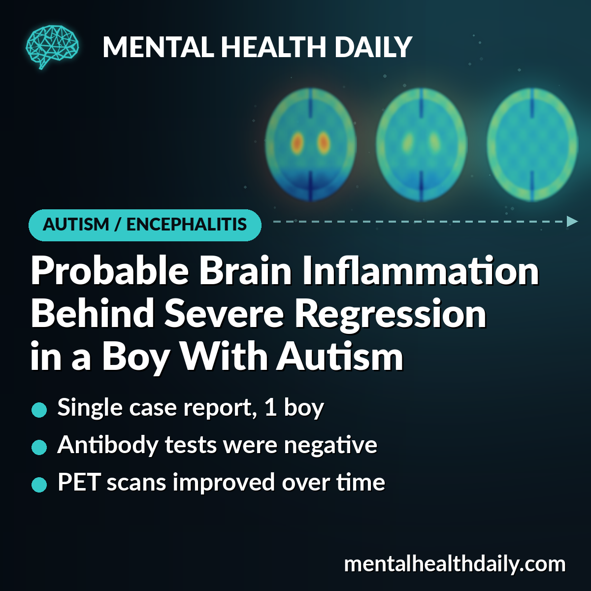
An 11-year-old boy with autism lost skills he had had for years, became aggressive, hurt himself many times a day, and did not get better with months of psychiatric treatment.
In a 2026 case report, doctors in Paris traced his decline to probable autoimmune encephalitis, a brain inflammation driven by the immune system, and used repeat brain scans to follow his partial recovery.1
Research Highlights
- One boy, one report: this is a single case, so it shows what can happen, not how often it happens.1
- Severe, unusual decline: aggression, self-injury, repetitive “behavioral loops,” psychotic-like behavior and loss of daily skills did not respond to several psychiatric drugs.1
- Clues beyond autism: seizure activity on EEG, changes in his spinal fluid and an abnormal brain PET scan pointed to an immune cause, even though antibody tests were negative.1
- Gradual, partial improvement: after monthly immune treatment, he improved step by step, and his last PET scan was within normal limits overall.1
- No single cause of recovery: he received immune treatment, clozapine, seizure medicines and rehabilitation together, so the report cannot say which one helped.1
How Did an 11-Year-Old Boy With Autism Lose His Skills?
Before the episode, the boy talked, was toilet trained, and attended a mainstream school with support. He had an autism diagnosis but no intellectual disability.1
From about September 2023, he declined over many months. He began wetting and soiling himself every day, refused to wear clothes, ate food off the floor, and pulled away from people.1
Early in this decline, he also had a liver inflammation linked to Epstein–Barr virus and parvovirus B19. It cleared up on its own in about 15 days.1
By the time he reached a specialized child psychiatry unit at Pitié-Salpêtrière Hospital in Paris, his symptoms included:1
- frequent aggression toward others and self-injury
- explosive outbursts and intense anxiety
- obsessive-compulsive-like “behavioral loops” and repeated words or phrases
- psychotic-like behavior, such as talking to himself, fixed staring and seeming to respond to things others could not see
- unusual movements of the jaw, tongue and right side of the body
The doctors described this as catatonic deterioration: a drop well below his usual baseline in movement, behavior and responsiveness. Antipsychotic, antidepressant, benzodiazepine and mood-stabilizer treatments had not brought lasting improvement.1
Why Doctors Looked Past Autism for an Explanation
When a child with autism gets much worse, it is easy to blame autism itself. Doctors call this diagnostic overshadowing: an existing diagnosis hides a new medical problem.1
Here, the sudden loss of skills, the unusual movements and the lack of response to standard treatment led the team to run a wide medical work-up.1
- EEG (brain wave recording): showed seizure-type discharges on the right side, consistent with focal epilepsy. After seizure medicines (valproate and lacosamide), later EEGs no longer showed them.
- MRI: no meaningful structural problem, apart from a small fluid-filled cyst found by chance.
- Spinal fluid: a first sample in December 2024 was contaminated with blood and hard to read. A repeat sample in September 2025 showed a mild rise in white blood cells and signs of a leaky barrier between blood and spinal fluid.
- Antibodies: tests for antibodies that attack brain cells were negative in both blood and spinal fluid.
- Other causes: tests for infection, lupus, thyroid disease, tumors, and metabolic and genetic conditions did not find another explanation.
Putting it together, the team settled on probable seronegative autoimmune encephalitis. “Seronegative” means the known brain-attacking antibodies were not found, so the diagnosis rests on the overall picture instead of a single positive test.1
What the Boy’s Brain PET Scans Showed Over 15 Months
An FDG-PET scan tracks how much sugar (glucose) different brain areas take up, a rough measure of how active they are.
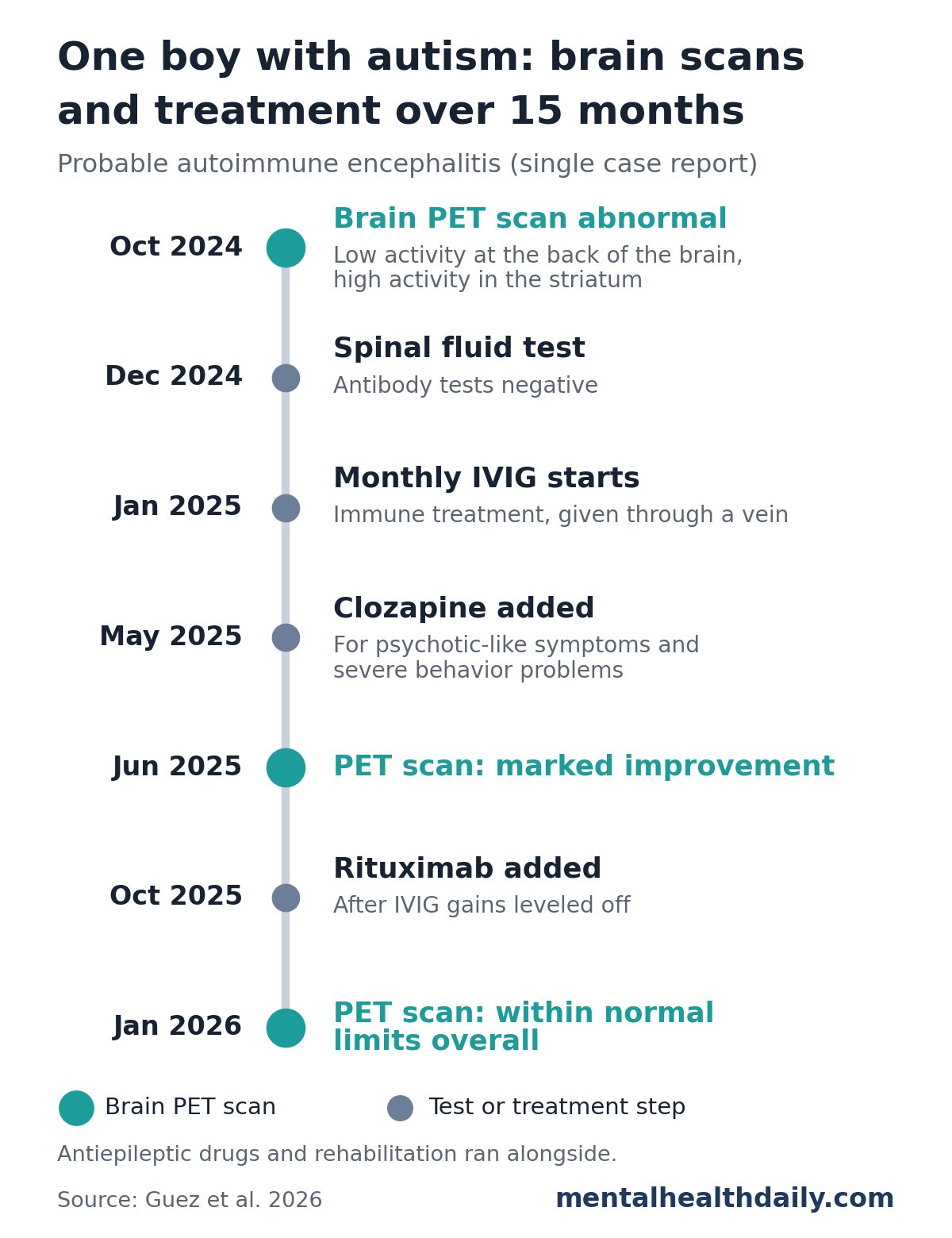
His first scan, in late October 2024, showed low activity in the back of the brain, mostly the occipital region that handles vision. It also showed high activity in the striatum, a deep structure involved in movement and habits, on both sides.1
A follow-up scan in late June 2025 showed marked improvement.
By late January 2026, the scan was reported as within normal limits overall. Striatal activity had normalized and the changes at the back of the brain were gone.1
The last scan had limits of its own. It was shorter than usual, blurred by movement, and still showed a mild, nonspecific dip in activity at the front of the brain.1
How the Boy Responded to IVIG, Rituximab and Clozapine
Once an immune cause was suspected, he started monthly IVIG (intravenous immunoglobulin, donated antibodies given through a vein to calm an overactive immune system) in January 2025.1
He did not respond fully right away after the first course. After the first 3 courses, his doctors estimated about 40% clinical improvement, but symptoms flared again before each next course.1
When his gains leveled off, he received rituximab, a drug that clears out B cells, the immune cells that go on to make antibodies, at the end of October 2025. He tolerated it well.1
Other treatment ran in parallel. Clozapine, an antipsychotic used when others fail, was added in late May 2025 and raised to 350 mg a day by late September 2025. He also stayed on seizure medicines and had structured rehabilitation and education.1
Staff counted his behavior incidents every week. At the worst points in autumn 2024, they recorded close to 800 self-injury incidents in a single week. By early 2026, weekly counts of self-injury, aggression and disruptive behavior were close to zero.1
That drop came with a change in setting: from January 2026, counts were recorded during twice-weekly day-hospital visits instead of full-time admission.
He became more emotionally available, regained some daily skills, and went on home visits and short hospital-based school sessions. He left full-time hospital care for day-hospital care twice a week. No severe infusion reactions, serious infections or neurological worsening were reported.1
Looking back, he told his parents that being in the hospital was hard, especially being apart from his family. He also described some staff as “firm but kind” and said, “this hospital treated me.”1
Why “Probable” Autoimmune Encephalitis and Not a Confirmed Diagnosis
Doctors who wrote the case report are careful about what one boy’s story can show. The main limits:
- Single case: one boy cannot tell us how common this is in children with autism, or who else might benefit from testing.
- Diagnosis not definite: under strict standard criteria, the case met only “possible” autoimmune encephalitis. MRI was normal, antibodies were negative, and the first spinal fluid sample was hard to interpret.
- Many treatments at once: IVIG, rituximab, clozapine, seizure medicines and rehabilitation all overlapped, so no one treatment can be credited.
- PET is not proof: the brain activity pattern is nonspecific. He also took calming medicines before the scans, including a benzodiazepine, and such drugs may affect PET readings.
- Partial recovery: he still had flare-ups around frustration, changes in routine, hygiene and treatment days.
This report offers no treatment advice for autism. Immune therapy here was given for suspected brain inflammation, not for autism itself.
What Earlier Research Says About Autoimmune Encephalitis in Autism
Standard criteria for autoimmune encephalitis, published in 2016, rank cases as possible, probable or definite. They were designed so doctors could act on clinical signs and routine tests instead of waiting on antibody results.2
A 2025 chart review of 67 children evaluated for autoimmune encephalitis included children with autism and other developmental conditions. Those diagnosed with it had symptoms across a similar number of areas as typically developing children, and a similar response to treatment.3
The same Paris group earlier reported a 16-year-old with probable seronegative encephalitis and catatonia, without autism. In that case, repeat PET scans helped with diagnosis, treatment decisions and monitoring, and the teen recovered after 2 years of treatment.4
Together, these studies support the main message of the Paris case report: when a child with autism has a severe, unusual decline that does not respond to standard care, an immune cause is worth checking, even when antibody tests are negative.1
References
- Guez E, Falempin T, Hanin C, et al. Serial FDG-PET in probable seronegative autoimmune encephalitis presenting as severe behavioral regression in a child with autism. Psychiatry and Clinical Neurosciences Reports. 2026;5:e70395. doi:10.1002/pcn5.70395
- Graus F, Titulaer MJ, Balu R, et al. A clinical approach to diagnosis of autoimmune encephalitis. Lancet Neurology. 2016;15(4):391–404. doi:10.1016/S1474-4422(15)00401-9
- Panzer K, Harmon A, Lerebours R, et al. Retrospective comparison of patients evaluated for pediatric autoimmune encephalitis with typical and atypical premorbid neuropsychiatric development. Journal of Autism and Developmental Disorders. 2025;55(6):2059–2066. doi:10.1007/s10803-023-06065-9
- Guetta M, Kas A, Aouidad A, et al. Relevance of brain 18F-FDG PET imaging in probable seronegative encephalitis with catatonia: a case report. Frontiers in Psychiatry. 2021;12:685711. doi:10.3389/fpsyt.2021.685711